. 2017 Jan 3;8(1):692-704.
doi: 10.18632/oncotarget.13524.

Cholesterol import and steroidogenesis are biosignatures for gastric cancer patient survival

Affiliations

Cholesterol import and steroidogenesis are biosignatures for gastric cancer patient survival

Wei-Chun Chang et al. Oncotarget. .

Abstract

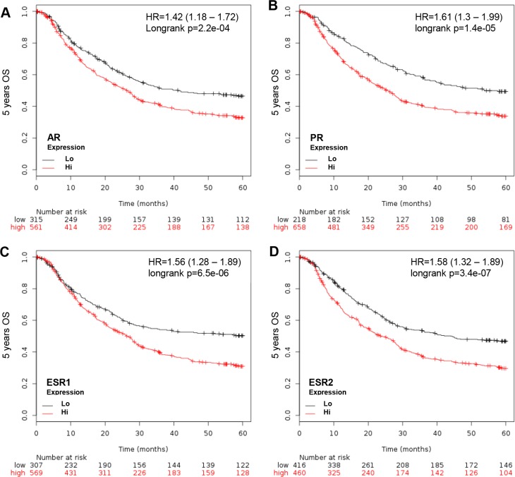
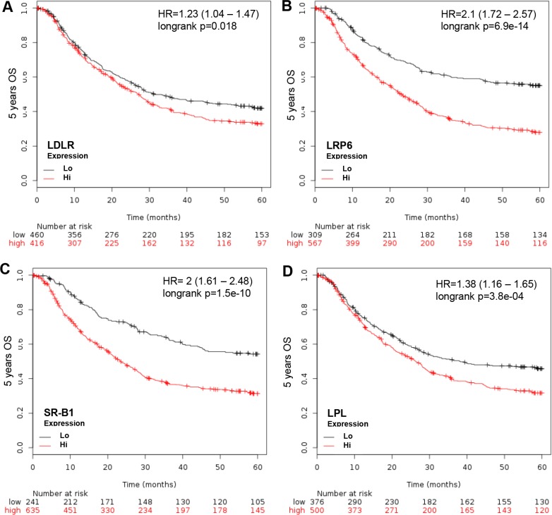
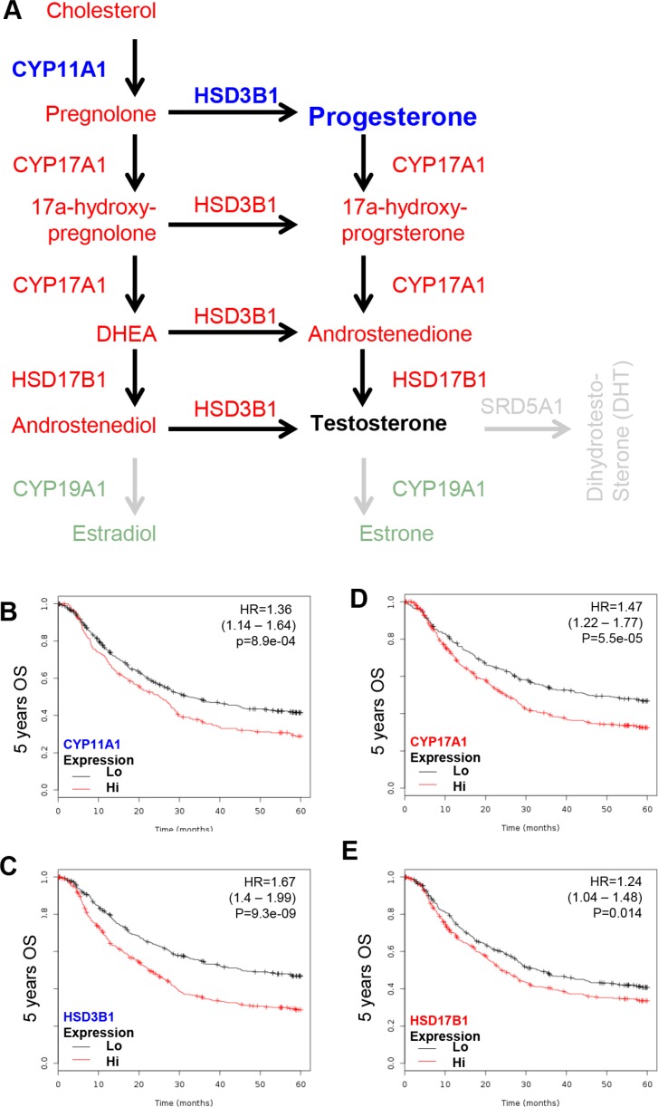
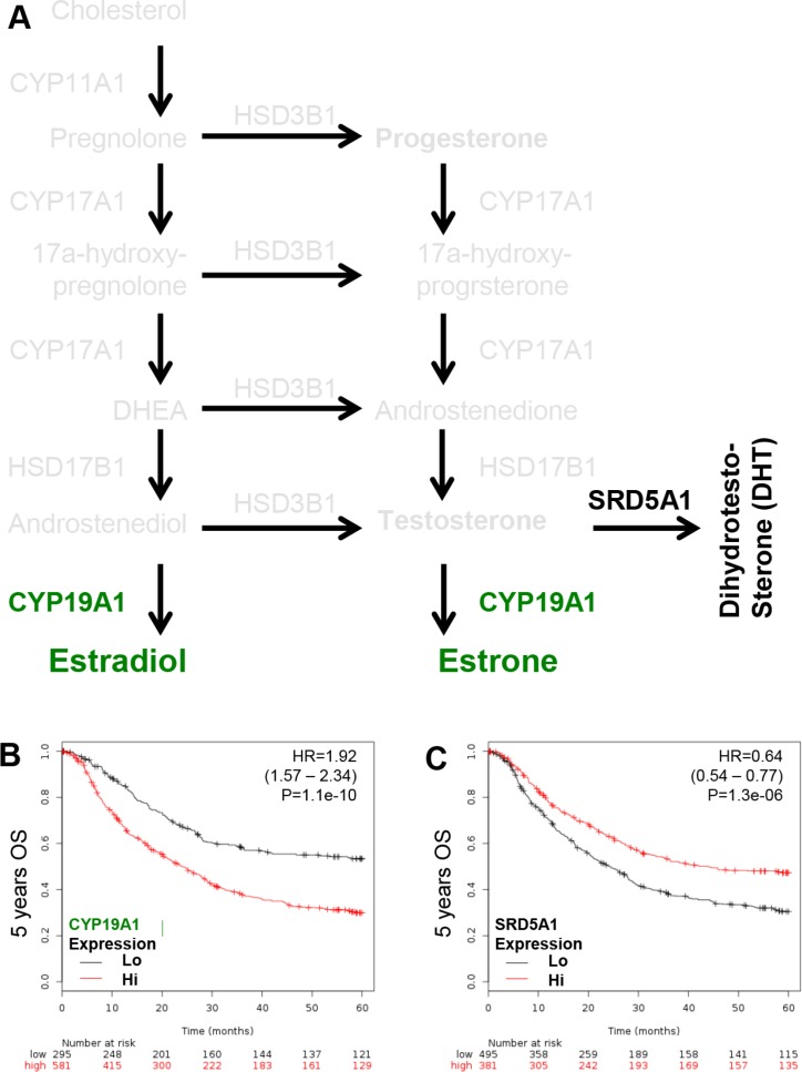
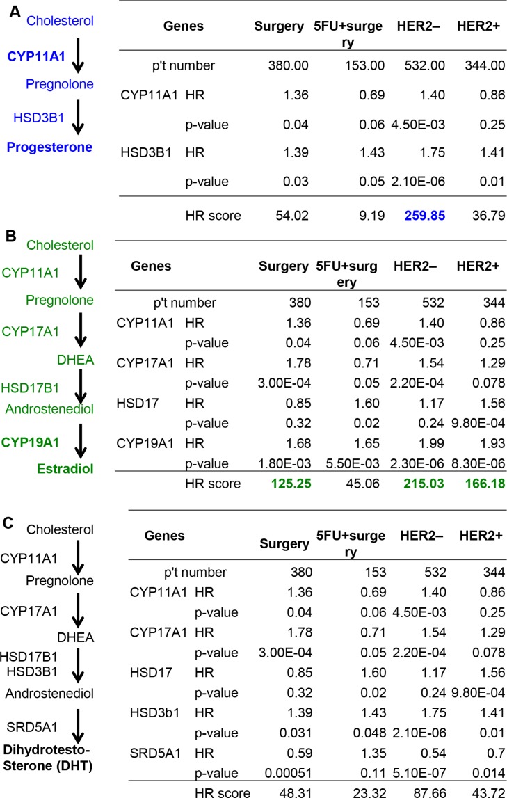
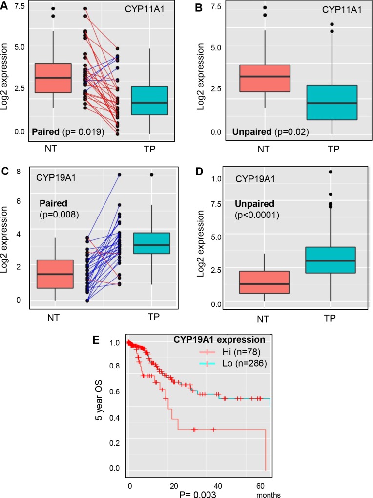
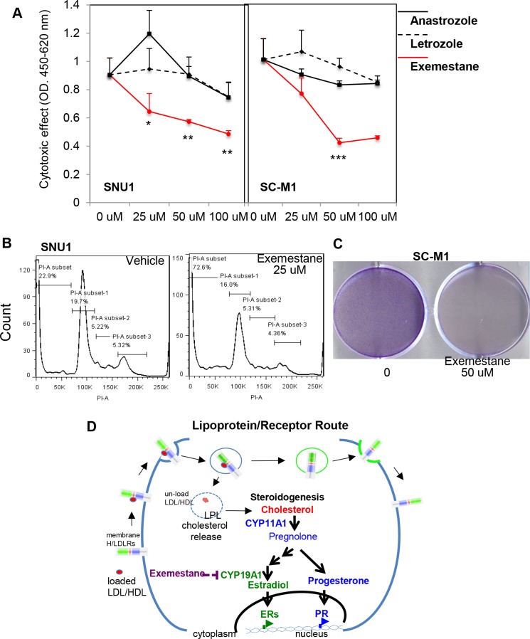
Androgens, estrogens, progesterone and related signals are reported to be involved in the pathology of gastric cancer. However, varied conclusions exist based on serum hormone levels, receptor expressions, and in vitro or in vivo studies. This report used a web-based gene survival analyzer to evaluate biochemical processes, including cholesterol importing via lipoprotein/receptors (L/R route), steroidogenic enzymes, and steroid receptors, in gastric cancer patients prognosis. The sex hormone receptors (androgen receptor, progesterone receptor, and estrogen receptor ESR1 or ESR2), L/R route (low/high-density lipoprotein receptors, LDLR/LRP6/SR-B1 and lipoprotein lipase, LPL) and steroidogenic enzymes (CYP11A1, HSD3B1, CYP17, HSD17B1, HSD3B1, CYP19A1 and SRD5A1) were associated with 5-year survival of gastric cancer patients. The AR, PR, ESR1 and ESR2 are progression promoters, as are the L/R route LDLR, LRP6, SR-B1 and LPL. It was found that CYP11A1, HSD3B1, CYP17, HSD17B1 and CYP19A1 promote progression, but dihydrotestosterone (DHT) converting enzyme SRD5A1 suppresses progression. Analyzing steroidogenic lipidome with a hazard ratio score algorithm found that CYP19A1 is the progression confounder in surgery, HER2 positive or negative patients. Finally, in the other patient cohort from TCGA, CYP19A1 was expressed higher in the tumor compared to that in normal counterparts, and also promoted progression. Lastly, exemestrane (type II aromatase inhibitor) dramatically suppress GCa cell growth in pharmacological tolerable doses in vitro. This work depicts a route-specific outside-in delivery of cholesterol to promote disease progression, implicating a host-to-tumor macroenvironmental regulation. The result indicating lipoprotein-mediated cholesterol entry and steroidogenesis are GCa progression biosignatures. And the exemestrane clinical trial in GCa patients of unmet medical needs is suggested.

Keywords: CYP19A1; cholesterol; gastric cancer; steroidogenesis.


Conflict of interest statement

CONFLICTS OF INTEREST

The US and Taiwan patents regarding the use of exemestrane for gatric cancer therapy have been filed out, and WL Ma is the inventor. Other authors claim no interest conflict in this work.

Figures

Figure 1
Figure 1. KM plotter evaluation of sex hormone nuclear receptors, including AR, PR, ESR1 and ESR2, in GCa 5-year OS
(A) AR expression status in GCa patients. Red line indicates high expression and black line indicates low expression. At the initial time point (0 months), 561 patients have high AR and 315 have low AR. At the last time point (60 months), 138 have high AR and 112 have low AR. The HR is 1.42 (range 1.18–1.72), and p-value is 2.2e-04. (B) PR expression status in GCa patients. The number of 0 month patients with high PR is 658, and low PR is 218. The 60-month patient number with high PR is 169, and low PR is 81. The HR is 1.61 (range 1.3–1.99), and p-value is 1.4e-05. (C) ESR1 expression status in GCa patients. The 0 month patient number with high ESR1 is 569, and low ESR1 is 307. The 60-month patient number of high ESR1 is 128, and low ESR1 is 122. The HR is 1.56 (range 1.28–1.89), and p-value is 6.5e-06. (D) ESR2 expression status in GCa patients. The 0 month patient number of high ESR2 is 460, and low ESR2 is 307. The 60–month patient number of high ESR2 is 104, and low ESR2 is 146. The HR is 1.58 (range 1.32–1.89), and p-value is 3.4e-07.
Figure 2
Figure 2. KM plotter evaluation of L/R route, including LDLR, LRP6, SR-B1 and LPL in GCa 5-year OS
(A) LDLR expression status in GCa patients. Red line indicates high expression and the black line indicates low expression. The 0 month patient number with high LDLR is 416, and low LDLR is 460. The last 60-month patient number with high LDLR is 97, and low LDLR is 153. The HR is 1.23 (range 1.04–1.47), and p-value is 0.018. (B) LRP6 expression status in GCa patients. The 0 month patient number with high LRP6 is 567, and low LRP6 is 309. The 60-month patient number with high LRP6 is 116, and low LRP6 is 134. The HR is 2.1 (range 1.72–2.57), and p-value is 6.9e-14. (C) SR-B1 expression status in GCa patients. The 0 month patient number with high SR-B1 is 639, and low SR-B1 is 241. The 60-month patient number with high SR-B1 is 145, and low SR-B1 is 105. The HR is 2 (range 1.61–2.48), and p-value is 1.5e-10. (D) LPL expression status in GCa patients. The 0 month patient number with high LPL is 500, and low LPL is 376. The 60-month patient number with high LPL is 120, and low LPL is 130. The HR is 1.38 (range 1.16–1.65), and p-value is 3.8e-04.
Figure 3
Figure 3. KM plotter evaluation of sex steroid lipidome related enzyme, including CYP11A1, CYP17, HSD3B1 and HSD17B1 in GCa 5-year OS
(A) Schematic illustration of sex steroid lipidomes and responding genes, including CYP11A1 (conversion of cholesterol to pregnolone; blue colored), CYP17 (conversion between pregnolone, 17a-hydroxyprognolone, androstenedione, progesterone, 17a-hydroxy- progesterone and DHEA; red colored), HSD3B1 (conversion of pregnolone, 17a-hydroxyprognolone, or androstenedione to progesterone, 17a-hydroxyprogesterone, or DHEA; red colored) and HSD17B1 (conversion of DHEA or androstenedione to androstenediol or testosterone; red colored). (B) CYP11A1 expression status in GCa patients. The HR is 1.36 (range 1.14–1.64), and p-value is 8.9e-04. (C) HSD3B1 expression status in GCa patients. The HR is 1.67 (range 1.4–1.99), and p-value is 9.3e-09. (D) CYP17 expression status in GCa patients. The HR is 1.47 (range 1.22–1.77), and p-value is 5.5e-05. (E) HSD17B1 expression status in GCa patients. The HR is 1.24 (range 1.04–1.48), and p-value is 0.014.
Figure 4
Figure 4. KM plotter evaluation of rate limiting step enzymes for estradiol (CYP19A1; aromatase) and DHT (SRD5A1; 5α-reductase) in GCa 5-years OS
(A) Schematic illustration of estradiol and DHT lipidomes and responding genes, including CYP19A1 (conversion of androstenedione or testosterone to estradiol or estrone; green colored), SRD5A1 (conversion of testosterone to DHT; black colored). (B) CYP19A1 expression status in GCa patients. The HR is 1.92 (range 1.57–2.34), and p-value is 1.1e-10. (C) SR-B1 expression status in GCa patients. The HR is 0.64 (range 0.54–0.77), and p-value is 1.3e-06.
Figure 5
Figure 5. Calculation of the HR score of steroidogenic lipidomes in GCa progression
(A) Progesterone anabolic enzymes CYP11A1 and HSD3B1 in surgery, 5-FU and surgery, HER2– or HER2+ subcategories of GCa patients. The HR score of each subcategory are: 54.02 for surgery, 9.19 for 5-FU and surgery, 259.85 for HER2– and 36.79 for HER2+ patients. (B) Estradiol anabolic enzymes CYP11A1, CYP17, HSD17B1 and CYP19A1 in the four subcategories of GCa patients. The HR score of each subcategory are: 125.25 for surgery, 45.06 for 5-FU and surgery, 215.03 for HER2– and 166.18 for HER2+ patients. (C) DHT anabolic enzymes CYP11A1, CYP17, HSD3B1 and SRD5A1 in four the subcategories of GCa patients. The HR score of each subcategory are: 48.31 for surgery, 23.32 for 5-FU and surgery, 87.66 for HER2– and 43.72 for HER2+ patients.
Figure 6
Figure 6. Expression analysis of CYP11A1 and CYP19A1 in non-tumor versus tumor parental lesions of GCa with DriverDB. v2 platform
(A) Paired comparison of CYP11A1 expression in non-tumor (NT) and tumor-parental (TP) of gastric cancer patients. TP is significantly lower than NT, with a p-value 0.019. (B) Unpaired comparison of CYP11A1 expressions in NT and TP of gastric cancer patients. TP is significantly lower than NT, with a p-value 0.02. (C) Paired comparison of CYP19A1 expressions in NT and TP of gastric cancer patients. TP is significantly higher than NT, with a p-value 0.008. (D) Unpaired comparison of CYP19A1 expressions in NT and TP of gastric cancer patients. TP is significantly higher than NT, with a p-value less than 0.0001. (E) Validation of the database cohort association of CYP19A1 expressions to 5-year overall survival (OS) in GCa patients from the TCGA database. The patients were divided to two groups: hi (high expression; n = 78), and lo (low expression; n = 286). The logrank test p-value = 0.003.
Figure 7
Figure 7. Targeting CYP19A1 with exemestane is novel therapy for GCa
(A) Cytotoxic analysis of CYP19A1 inhibitors (Anastrazole, Letrozole, and Exemestane) in SNU1 (left panel) and SC-M1 (right panel) cells. The cytotoxic effect can be observed with exmestane, but not anastrazole and letrozole treatments. (B) Apoptotic cells was dramatically increased in SNU1 cells in 25 μM exemestane (right panel) compared with vehicle (left panel) treatments. (C) Long-term exemestane (25 μM; 7-days) can totally suppresses colony formation in SC-M1 cells compared to vehicle treatments. (D) Schematic illustration of L/R route to steroidogenesis in GCa cells. The cell membrane L/R route complex (LDLR, LRP6, SR-B1 and LPL) shuttles extracellular cholesterol carriers (e.g., LDL or HDL) into cancer cells. LPL is the catalysis to release cholesterol into cells, and receptors are then recycled back to the cell membrane. Increased cellular cholesterol provides resources for steroidogenesis, particularly CYP11A to increase progesterone or CYP19A1 to increase estradiol for PR or ESRs, respectively. Activated receptors could then alter genome-wide transcriptomes to promote cancer progression. Targeting steroidogenesis, e.g., CYP19A1 by Aromatase Inhibitors, exemestane, might be an effective therapeutic regimen in GCa patients.

References

    1. Thrumurthy SG, Chaudry MA, Chau I, Allum W. Does surgery have a role in managing incurable gastric cancer? Nat Rev Clin Oncol. 2015;12:676–82. - PubMed
    1. Tan P, Yeoh KG. Genetics and Molecular Pathogenesis of Gastric Adenocarcinoma. Gastroenterology. 2015;149:1153–62. e3. - PubMed
    1. Bang YJ, Van Cutsem E, Feyereislova A, Chung HC, Shen L, Sawaki A, Lordick F, Ohtsu A, Omuro Y, Satoh T, Aprile G, Kulikov E, Hill J, et al. Trastuzumab in combination with chemotherapy versus chemotherapy alone for treatment of HER2-positive advanced gastric or gastro-oesophageal junction cancer (ToGA): a phase 3, open-label, randomised controlled trial. Lancet. 2010;376:687–97. - PubMed
    1. Allum WH, Blazeby JM, Griffin SM, Cunningham D, Jankowski JA, Wong R. Guidelines for the management of oesophageal and gastric cancer. Gut. 2011;60:1449–72. - PubMed
    1. Foo M, Leong T. Adjuvant therapy for gastric cancer: current and future directions. World J Gastroenterol. 2014;20:13718–27. - PMC - PubMed

MeSH terms