. 2016 Nov 28;10(11):e0005102.
doi: 10.1371/journal.pntd.0005102. eCollection 2016 Nov.

Molecular Epidemiology of Agents of Human Chromoblastomycosis in Brazil with the Description of Two Novel Species

Affiliations

Molecular Epidemiology of Agents of Human Chromoblastomycosis in Brazil with the Description of Two Novel Species

Renata R Gomes et al. PLoS Negl Trop Dis. .

Erratum in

Abstract

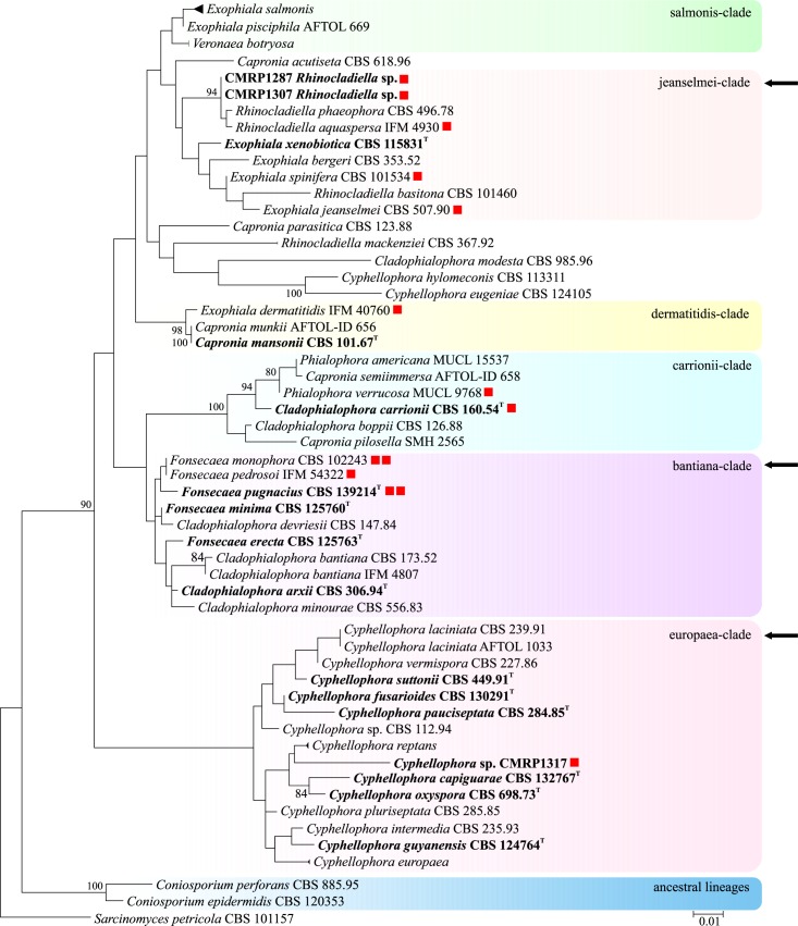
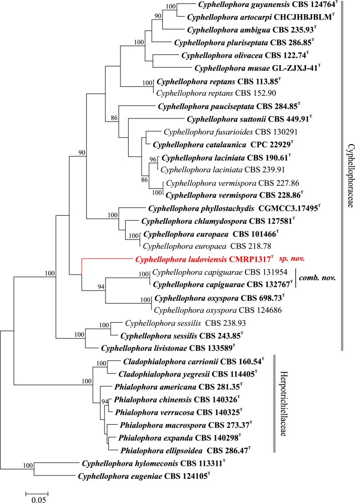
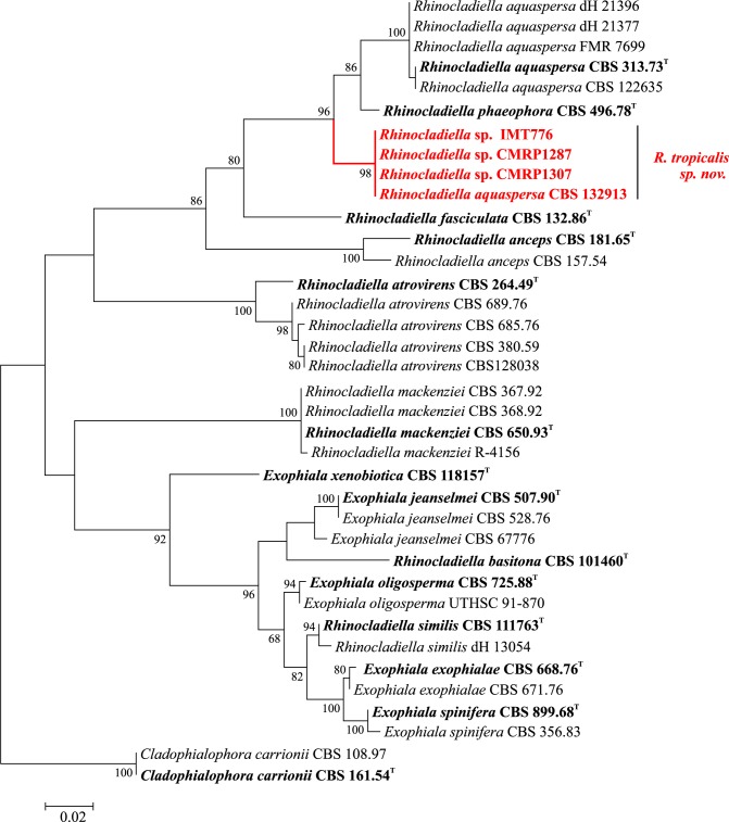
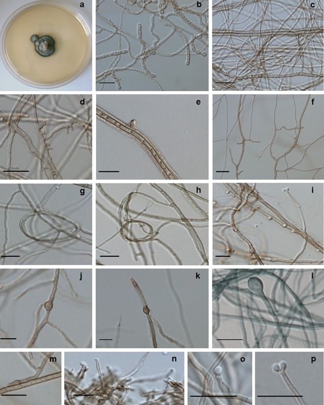
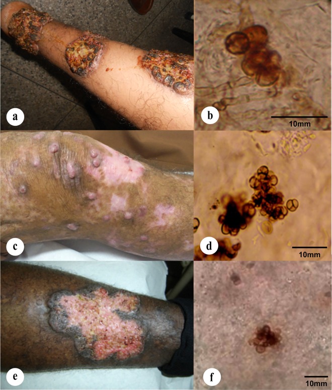
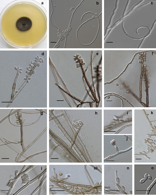
The human mutilating disease chromoblastomycosis is caused by melanized members of the order Chaetothyriales. To assess population diversity among 123 clinical strains of agents of the disease in Brazil we applied sequencing of the rDNA internal transcribed spacer region, and partial cell division cycle and β-tubulin genes. Strains studied were limited to three clusters divided over the single family Herpotrichiellaceae known to comprise agents of the disease. A Fonsecaea cluster contained the most important agents, among which F. pedrosoi was prevalent with 80% of the total set of strains, followed by 13% for F. monophora, 3% for F. nubica, and a single isolate of F. pugnacius. Additional agents, among which two novel species, were located among members of the genus Rhinocladiella and Cyphellophora, with frequencies of 3% and 1%, respectively.


Conflict of interest statement

The authors have declared that no competing interests exist.

Figures

Fig 1
Fig 1. Phylogeny of a representative selection of species in Chaetothyriales, based on confidently aligned LSU sequences.
Constructed with Maximum likelihood implemented in MEGA 7. Bootstrap values > 80% from 100 resampled datasets are shown with branches. Coloured boxes represent species complexes taken from de Hoog et al. [21], Feng et al. [22], and Vicente et al. [23]. Clades with species causing chromoblastomycosis analysed in this study are indicated with arrows. Type strain in bold.
Fig 2
Fig 2. Multilocus tree of Cyphellophora based on ITS and partial BT2 sequences.
Constructed with maximum likelihood implemented in MEGA 7. Bootstrap values of >80% from 100 resampled data sets are shown with branches. Cladophialophora yegresii and C. carrionii comprised the outgroup. Novel species causing chromoblastomycosis are indicated with red branches. Type strain in bold.
Fig 3
Fig 3. Multilocus tree of Rhinocladiella based on ITS and partial BT2 sequences.
Constructed with maximum likelihood implemented in MEGA 7. Bootstrap values of >80% from 100 resampled data sets are shown with branches. Cladophialophora yegresii and C. carrionii comprised the outgroup. Novel species causing chromoblastomycosis are indicated with red branches. Type strain in bold.
Fig 4
Fig 4. Cardinal temperatures of strains described.
(A) Cyphellophora ludoviensis with optimal growth temperature at 30°C and maximum at 37°C. (B) Rhinocladiella tropicalis with optimal development at 27°C and maximum at 37°C.
Fig 5
Fig 5. Cyphellophora ludoviensis microscopic morphology.
(A) colonies on SGA; (B-E) hyphae with chlamydospores and lateral extensions; (F) anastomosis; (G-H) spirally twisted hyphae; (I) poorly differentiated phialide producing conidia; (J-P) chlamydospores and conidia. Scale bars 10 μm.
Fig 6
Fig 6. Clinical case pictures.
(A, B) Nodular and verrucous lesions and muriform cells from skin tissue biopsy of arm lesion caused by Cyphellophora ludoviensis (CMRP1317); (C-E) polymorphic infiltrative plaque lesions caused by different strains of Rhinocladiella tropicalis affecting the legs, (C) with nodular and cicatricial and (E) verrucous lesions; (D-F) muriform cells from skin tissue biopsy of lesions caused by R. tropicalis strains CMRP1287 and CMRP1307, respectively.
Fig 7
Fig 7. Rhinocladiella tropicalis, microscopic morphology.
(A) Colonies on SGA; (B, C) twisted hyphae and conidia; (D-G) conidiophores with conidia produced in sympodial order; (H-O) conidial apparatus with conidia. Scale bars 10 μm.

References

    1. Deng S, Tsui CKM, Gerrits van den Ende AHG, Yang L, Najafzadeh M J, Badali H, et al. (2015) Global spread of human chromoblastomycosis is driven by recombinant Cladophialophora carrionii and predominantly clonal Fonsecaea species. PLoS Negl Trop Dis 10: 1–15. - PMC - PubMed
    1. Queiroz-Telles F, Santos DW (2013) Challenges in the therapy of chromoblastomycosis. Mycopathologia. 175: 477–488. 10.1007/s11046-013-9648-x - DOI - PubMed
    1. Silva JP, de Souza W, Rozental S (1998) Chromoblastomycosis: a retrospective study of 325 cases on Amazonic Region (Brazil). Mycopathologia 143:171–175. - PubMed
    1. Bayles MA (1986) Chromomycosis In Hay RJ (ed.), Baillie`re’s Clinical Tropical Medicine and Comunicable Diseases. Tropical Fungal Infections. London: WB Saunders, 1986: 45–70.
    1. Esterre P, Pecarrère JL, Raharisolo C, Huerre M (1999) Squamous cell carcinoma arising from chromomycosis. Report of two cases. Ann Pathol 19:516–520. - PubMed

Publication types

LinkOut - more resources