. 2016 Dec 27;7(52):87147-87160.
doi: 10.18632/oncotarget.13515.

Negative feedback between TAp63 and Mir-133b mediates colorectal cancer suppression

Affiliations

Negative feedback between TAp63 and Mir-133b mediates colorectal cancer suppression

Jing Dai et al. Oncotarget. .

Abstract

Background: TAp63 is known as the most potent transcription activator and tumor suppressor. microRNAs (miRNAs) are increasingly recognized as essential components of the p63 pathway, mediating downstream post-transcriptional gene repression. The aim of present study was to investigate a negative feedback loop between TAp63 and miR-133b.

Results: Overexpression of TAp63 inhibited HCT-116 cell proliferation, apoptosis and invasion via miR-133b. Accordingly, miR-133b inhibited TAp63 expression through RhoA and its downstream pathways. Moreover, we demonstrated that TAp63/miR-133b could inhibit colorectal cancer proliferation and metastasis in vivo and vitro.

Materials and methods: We evaluated the correlation between TAp63 and miR-133b in HCT-116 cells and investigated the roles of the TAp63/miR-133b feedback loop in cell proliferation, apoptosis and metastasis via MTT, flow cytometry, Transwell, and nude mouse xenograft experiments. The expression of TAp63, miR-133b, RhoA, α-tubulin and Akt was assessed via qRT-PCR, western blot and immunofluorescence analyses. miR-133b target genes were identified through luciferase reporter assays.

Conclusions: miR-133b plays an important role in the anti-tumor effects of TAp63 in colorectal cancer. miR-133b may represent a tiemolecule between TAp63 and RhoA, forming a TAp63/miR-133b/RhoA negative feedback loop, which could significantly inhibit proliferation, apoptosis and metastasis.

Keywords: TAp63; metastasis; miR-133b; negative feedback; proliferation.


Conflict of interest statement

CONFLICTS OF INTEREST

The authors declare no conflicts of interest.

Figures

Figure 1
Figure 1. Overexpression of TAp63 inhibits cell proliferation, apoptosis and microtubule formation
HCT-116 cells were transfected with the TAp63-expressing vector or negative controls (NCs). The expression of TAp63 was analyzed via qRT–PCR A. and western blotting B. C. The growth curves of the cells transfected with the TAp63-expressing vector or NCs were compared using the MTT assay. *P < 0.05. D. Distribution of cells in three phases (G1, S, and G2) of the cell cycle, as determined by flow cytometry analysis. Cytometric quantification of the experiments described in the chart, showing the proportions of cells in G1, S, and G2 phases.*P < 0.05. E. The apoptosis assay shows that the apoptosis rate in HCT-116/TAp63 cells was significantly higher than in the control. *P < 0.05.
Figure 2
Figure 2. TAp63 regulates microtubule formation and invasion
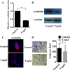
HCT-116 cells were transfected with the TAp63-expressing vector or negative controls (NCs). The expression of α-tubulin was analyzed via A. qRT–PCR, B. western blotting and C. immunofluorescence. D. Transwell invasion assays of HCT-116 and SW-620 cells transfected with either TAp63 or NC. After 48 h, invasive cells were counted in five random high-power fields. All of the data represent the means±sd of three different experiments analyzed in triplicate. *P<0.05.
Figure 3
Figure 3. TAp63 inhibits cell proliferation through miR-133b
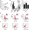
HCT-116 cells were transfected with the miR-133b-expressing vector or the negative controls (NCs), or co-transfected with the TAp63-expressing vector and miR-133b sponge. A. The growth curves of the cells transfected with the miR-133b-expressing vector or NCs, or co-transfected with the TAp63-expressing vector and miR-133b sponge were compared using the MTT assay. *P < 0.05. B. Cytometric quantification of the cell cycle analysis experiments described in the chart, showing the proportions of cells in G1, S, and G2 phases.*P < 0.05. C. Apoptosis rates determined via apoptosis assays in HCT-116 cells transfected with the miR-133b-expressing vector or NC, or co-transfected with the TAp63-expressing vector and miR-133b sponge TAp63+sponge. *P < 0.05, #P>0.05. D. Distribution of cells in three phases (G1, S, and G2) of the cell cycle, as determined by flow cytometry analysis. E. Apoptosis was measured via FACS.
Figure 4
Figure 4. TAp63 regulates microtubule formation and invasion through miR-133b
HCT-116 cells were transfected with the miR-133b-expressing vector or negative controls (NCs), or co-transfected with the TAp63-expressing vector and miR-133b sponge (TAp63+sponge). The expression of α-tubulin was analyzed via A. qRT–PCR, B. western blotting and C. immunofluorescence, *P < 0.05, #P>0.05. D. Transwell invasion assays of HCT-116 and SW-620 cells transfected with either TAp63 or NC. After 48 h, invasive cells were counted in five random high-power fields. All of the data represent the means±sd of three different experiments analyzed in triplicate. *P < 0.05, #P>0.05.
Figure 5
Figure 5. miR-133b regulates TAp63 expression
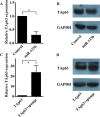
A. qRT-PCR was performed to detect the expression levels of TAp63 following transfection with the miR-133b-expressing vector or negative controls in HCT-116 cells. The data are expressed as the means±sd. *P<0.05. B. Western blotting was performed to detect the protein expression levels of TAp63 following transfection with the miR-133b-expressing vector or negative controls in HCT-116 cells. C. qRT-PCR was performed to detect the expression levels of TAp63 following transfection with the TAp63-expressing vector or co-transfection with the TAp63-expressing vector and miR-133b sponge (TAp63+sponge) in HCT-116 cells. The data are expressed as the means±sd. *P<0.05. D. Western blotting was performed to detect the protein expression levels of TAp63 following transfection with the TAp63-expressing vector or co-transfection with the TAp63-expressing vector and miR-133b sponge (TAp63+sponge) in HCT-116 cells.
Figure 6
Figure 6. miR-133b regulates TAp63 expression through the RhoA and PI3K/Akt pathways
A. qRT-PCR analysis of RhoA in HCT-116 cells transfected with the miR-133b-expressing vector or negative controls. The data are expressed as the means±sd. *P<0.05. B. Western blot analysis of RhoA and p-PI3K in HCT-116 cells transfected with the miR-133b-expressing vector or negative controls. C. qRT-PCR analysis of TAp63 in HCT-116 cells transfected with the negative controls or co-transfected with the miR-133b- and RhoA-expressing vectors. The data are expressed as the means±sd. #P>0.05. D. Western blot analysis of TAp63 and p-PI3K in HCT-116 cells transfected with the negative controls or co-transfected with the miR-133b- and RhoA-expressing vectors. E. qRT-PCR analysis of TAp63 in HCT-116 cells transfected with RhoA siRNA or the negative controls. The data are expressed as the means±sd. *P<0.05. F. Western blot analysis of TAp63 and p-PI3K in HCT-116 cells transfected with RhoA siRNA or the negative controls.
Figure 7
Figure 7. TAp63/miR-133b inhibits tumorigenicity in vivo
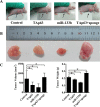
A. Tumors formed in nude mice. HCT-116 cells stably transfected with the TAp63-expressing vector, miR-133b-expressing vector or negative controls (NCs) or co-transfected with the TAp63-expressing vector with miR-133b sponge (TAp63+sponge) were injected into the flanks of nude mice (n=5), and the mice were sacrificed after 4 weeks. B. Images of whole tumors from nude mice injected with HCT-116 cells stably transfected with the TAp63-expressing vector, miR-133b-expressing vector or negative controls (NC), or co-transfected with the TAp63-expressing vector and miR-133b sponge (TAp63+sponge). C. The average volume and weight of the tumors from the mice injected with HCT-116 cells stably transfected with the TAp63-expressing vector or the miR-133b-expressing vector were significantly lower than that in those injected with negative control cells. The data are expressed as the means±sd. *P<0.05.
Figure 8
Figure 8. TAp63/miR-133b inhibits lung and liver metastasis
A. Representative gross lung images from nude mice injected intravenously with HCT-116 cells stably transfected with the TAp63-expressing vector, miR-133b-expressing vector or negative controls (NC), or co-transfected with the TAp63-expressing vector and miR-133b sponge (TAp63+sponge) through the tail vein are shown, with metastases visible at the lung surface marked with bold black arrows. B. Liver tissue levels of TAp63, miR-133b and RhoA were measured via qRT-PCR in nude mice injected intravenously with HCT-116 cells stably transfected with the TAp63-expressing vector, miR-133b-expressing vector or negative controls (NC), or co-transfected with the TAp63-expressing vector and miR-133b sponge (TAp63+sponge) through the tail vein. *P<0.05.

Similar articles

Cited by

References

    1. Celardo I, Grespi F, Antonov A, Bernassola F, Garabadgiu AV, Melino G, Amelio I. Caspase-1 is a novel target of p63 in tumor suppression. Cell Death Dis. 2013;4:e645. - PMC - PubMed
    1. Moll UM, Slade N. p63 and p73: roles in development and tumor formation. Mol Cancer Res. 2004;2:371–386. - PubMed
    1. Fischer B, Metzger M, Richardson R, Knyphausen P, Ramezani T, Franzen R, Schmelzer E, Bloch W, Carney TJ, Hammerschmidt M. p53 and TAp63 promote keratinocyte proliferation and differentiation in breeding tubercles of the zebrafish. PLoS Genet. 2014;10:e1004048. - PMC - PubMed
    1. Hershkovitz Rokah O, Shpilberg O, Granot G. NAD(P)H quinone oxidoreductase protects TAp63gamma from proteasomal degradation and regulates TAp63gamma-dependent growth arrest. PLoS One. 2010;5:e11401. - PMC - PubMed
    1. Yamaki T, Suenaga Y, Iuchi T, Alagu J, Takatori A, Itami M, Araki A, Ohira M, Inoue M, Kageyama H, Yokoi S, Saeki N, Nakagawara A. Temozolomide suppresses MYC via activation of TAp63 to inhibit progression of human glioblastoma. Sci Rep. 2013;3:1160. - PMC - PubMed