

Human limb skeletal muscle wasting and architectural remodeling during five to ten days intubation and ventilation in critical care - an observational study using ultrasound
- PMID: 27894277
- PMCID: PMC5127036
- DOI: 10.1186/s12871-016-0269-z
Human limb skeletal muscle wasting and architectural remodeling during five to ten days intubation and ventilation in critical care - an observational study using ultrasound
Abstract
Background: Critically ill patients frequently suffer muscle weakness whilst in critical care. Ultrasound can reliably track loss of muscle size, but also quantifies the arrangement of the muscle fascicles, known as the muscle architecture. We sought to measure both pennation angle and fascicle length, as well as tracking changes in muscle thickness in a population of critically ill patients.
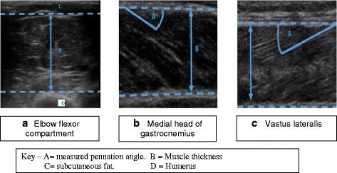
Methods: On days 1, 5 and 10 after admission to critical care, muscle thickness was measured in ventilated critically ill patients using bedside ultrasound. Elbow flexor compartment, medial head of gastrocnemius and vastus lateralis muscle were investigated. In the lower limb, we determined the pennation angle to derive the fascicle length.
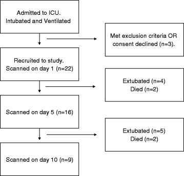
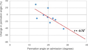
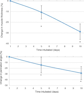
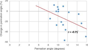
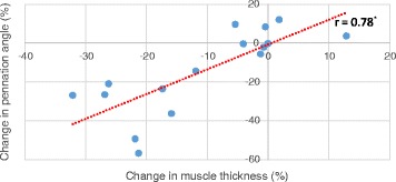
Results: We recruited and scanned 22 patients on day 1 after admission to critical care, 16 were re-scanned on day 5 and 9 on day 10. We found no changes to the size of the elbow flexor compartment over 10 days of admission. In the gastrocnemius, there were no significant changes to muscle thickness or pennation angle over 5 or 10 days. In the vastus lateralis, we found significant losses in both muscle thickness and pennation angle on day 5, but found that fascicle length is unchanged. Loss of muscle on day 5 was related to decreases in pennation angle. In both lower limb muscles, a positive relationship was observed between the pennation angle on day 1, and the percentage of angle lost by days 5 and 10.
Discussion: Muscle loss in critically ill patients preferentially affects the lower limb, possibly due to the lower limb becoming prone to disuse atrophy. Muscle architecture of the thigh changes in the first 5 days of admission, in particular, we have demonstrated a correlation between muscle thickness and pennation angle. It is hypothesised that weakness in the lower limb occurs through loss of force generation via a reduced pennation angle.
Conclusion: Using ultrasound, we have been able to demonstrate that muscle thickness and architecture of vastus lateralis undergo rapid changes during the early phase of admission to a critical care environment.
Keywords: Critical care; Muscle architecture; Muscle wasting; Pennation angle; Ultrasound; Weakness.
Figures
References
Publication types
MeSH terms
LinkOut - more resources
Full Text Sources
Other Literature Sources
Medical
