Review
. 2017 Jan:44:35-82.
doi: 10.1016/j.yfrne.2016.11.003. Epub 2016 Nov 25.

The dynamics of GABA signaling: Revelations from the circadian pacemaker in the suprachiasmatic nucleus

Affiliations
Review

The dynamics of GABA signaling: Revelations from the circadian pacemaker in the suprachiasmatic nucleus

H Elliott Albers et al. Front Neuroendocrinol. 2017 Jan.

Abstract

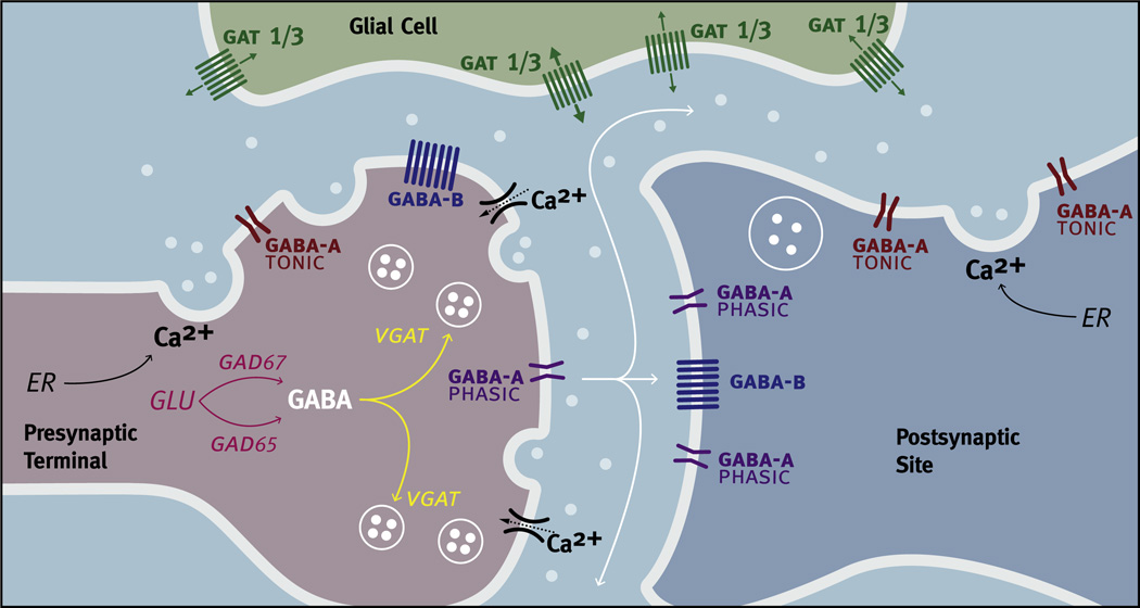
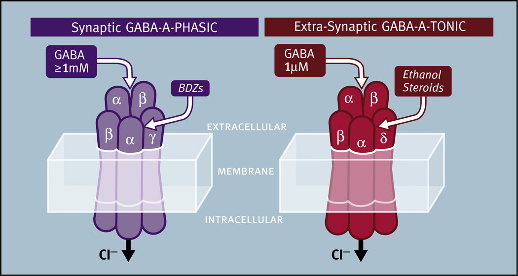
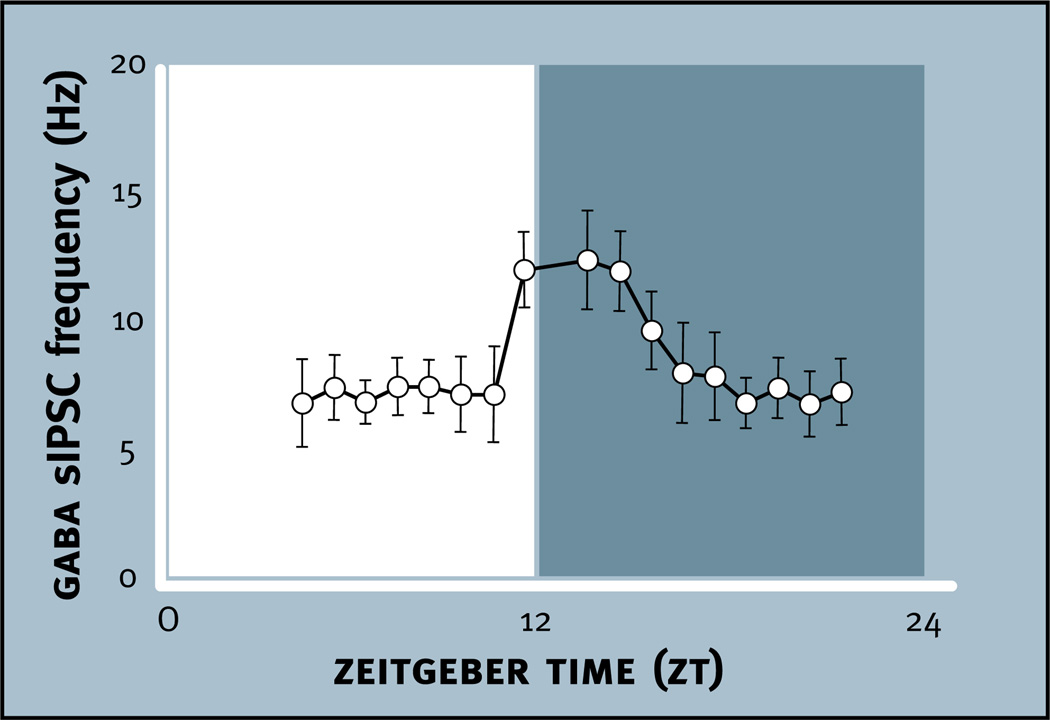
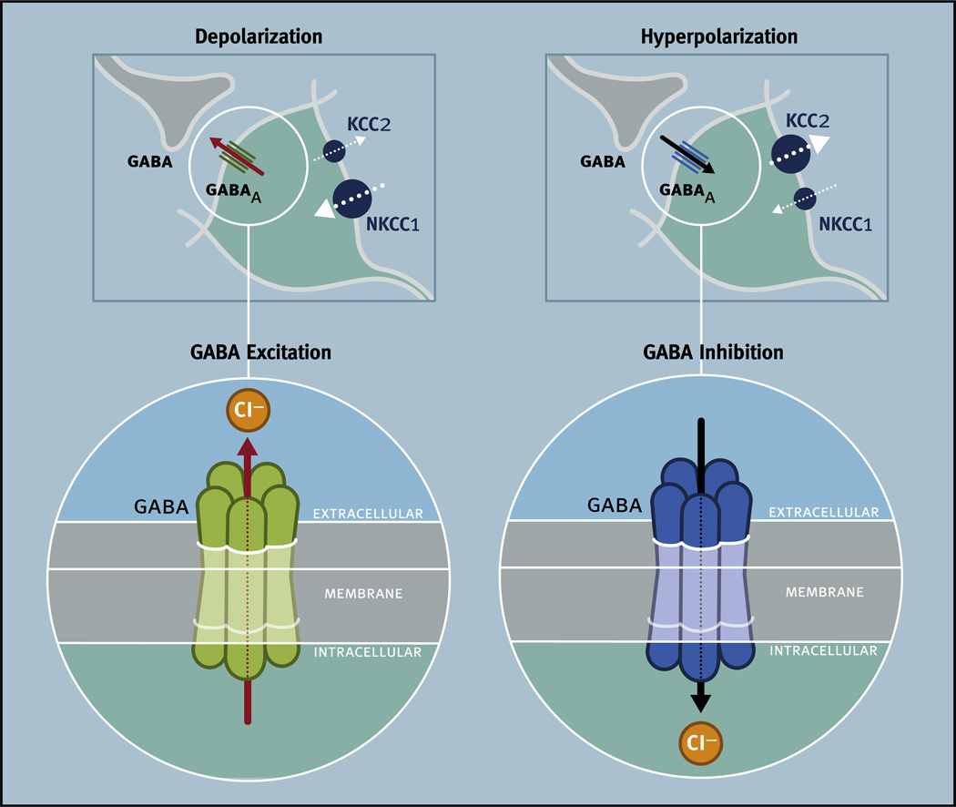
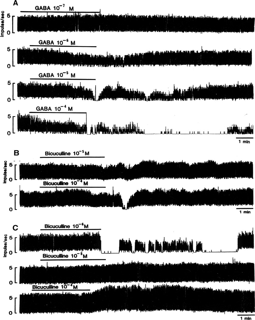
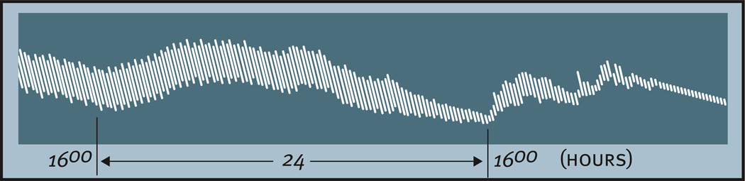
Virtually every neuron within the suprachiasmatic nucleus (SCN) communicates via GABAergic signaling. The extracellular levels of GABA within the SCN are determined by a complex interaction of synthesis and transport, as well as synaptic and non-synaptic release. The response to GABA is mediated by GABAA receptors that respond to both phasic and tonic GABA release and that can produce excitatory as well as inhibitory cellular responses. GABA also influences circadian control through the exclusively inhibitory effects of GABAB receptors. Both GABA and neuropeptide signaling occur within the SCN, although the functional consequences of the interactions of these signals are not well understood. This review considers the role of GABA in the circadian pacemaker, in the mechanisms responsible for the generation of circadian rhythms, in the ability of non-photic stimuli to reset the phase of the pacemaker, and in the ability of the day-night cycle to entrain the pacemaker.

Keywords: Benzodiazepines; Cation chloride cotransporters; Entrainment; Ethanol; GABA vesicular transporters; GABA(A) receptors; GABA(B) receptors; Glutamic acid decarboxylase; Membrane GABA transporters; Neurosteroids.


Figures

Fig. 1
Fig. 1
Comparison of the phase shifting effects of “photic” (solid red line) and “non-photic” stimuli (dotted black line) presented to nocturnally active rodents housed in constant darkness. Light does not produce phase shifts until the late subjective day and early subjective night when it produces phase delays. Later in the subjective night light produces phase advances. Non-photic stimuli, such as injection of neuropeptide Y into the suprachiasmatic region, induce large phase advances during the subjective day and smaller phase delays in the subjective night. Note: not all “non-photic” phase shifting stimuli produce a pattern of phase shifts like those seen in this figure. In nocturnal rodents the subjective day refers to the inactive phase and the subjective night refers to the active phase of the circadian cycle. Circadian time 12 is designated as the time of locomotor onset (modified from Webb et al., 2014).
Fig. 2
Fig. 2
Schematic diagram of the ventral core/dorsal shell conceptualization of the organization of the suprachiasmatic nucleus illustrating the anatomical and functional heterogeneity of the nucleus. Major afferent pathways including the retinohypothalamic tract (RHT), geniculohypothalamic tract (GHT), and a projection from the raphe nucleus (Raphe) terminate primarily in the ventral core, although RHT terminals can also be found in the dorsal shell. Neurons in the ventral core contain GABA and a variety of neuropeptides including vasoactive intestinal peptide (VIP) and gastrin releasing peptide (GRP), all of which are frequently colocalized in the same neuron. Some neurons in the ventral core display endogenous rhythmicity (~) while others do not (−). In the dorsal shell, more neurons display endogenous rhythmicity. Neurons in the dorsal shell contain GABA and neuropeptides including arginine-vasopressin (AVP), all of which are frequently colocalized in the same neuron. Neurons in the dorsal shell and ventral core can communicate via GABA and probably other neurochemical signals.
Fig. 3
Fig. 3
Hypothetical illustration of how different patterns of synaptic release of an amino acid neurotransmitter (NT) and colocalized neuropeptides (NP) could result from differences in neuronal firing and neuropeptide biosynthesis. Each panel is an example of differences that might occur in NT and NP release at a specific phase of the circadian cycle. (A) Moderate neuronal firing (l l l l l l) produces moderate levels of Ca2+ influx through voltage-gated ion channels resulting in exocytosis of small synaptic vesicles (SSV; purple circles) and release of NT. (B) High levels of neuronal firing (lllllllllll) produce high levels of Ca2+ influx through voltage-gated ion channels resulting in exocytosis of both SSVs and large dense-core vesicles (LDCV; blue circles) resulting in release of NT and NP. (C) Two different neuropeptides are packaged in LDCVs (red and blue) in a ratio of 1:1. High levels of neuronal firing (lllllllllll) produce high levels of Ca2+ influx through voltage-gated ion channels, resulting in exocytosis of SSVs and release of NT and exocytosis of LDCVs and release of a “cocktail” of neuropeptides in a 1:1 ratio. (D) Two different neuropeptides are packaged in LDCVs (red and blue) in a ratio of 3:1. High levels of neuronal firing (lllllllllll) produce high levels of Ca2+ influx through voltage-gated ion channels resulting in exocytosis of SSVs and release of NT and exocytosis of LDCVs and release of a “cocktail” of neuropeptides in a 3:1 ratio. Differential regulation of the biosynthesis and storage of neuropeptides could result in different ratios of neuropeptide release. If neuropeptide biosynthesis is differentially regulated over the circadian cycle then different ratios of neuropeptide would be released at different times of day (modified from Albers (2015)).
Fig. 4
Fig. 4
Factors regulating GABA signaling. Glutamic acid decarboxylase (GAD) catalyzes glutamic acid (GLU) into GABA in presynaptic neurons (light red). There are two isoforms of GAD. GAD67 synthesizes GABA for tonic release, while GAD65 synthesizes GABA for phasic release. GABA vesicular transporters (VGATs) are responsible for the transport of GABA into synaptic vesicles (yellow). GABA (white circles) can be found both in small synaptic vesicles (SSVs) and in large dense core-vesicles (LDCVs). The exocytosis of SSVs and LDCVs occurs in response to increases in intracellular calcium (Ca2+) resulting in the release of GABA into the extracellular space. Increases in intracellular Ca2+ can result from the influx of Ca2+ through voltage-gated ion channels as the result of an action potential or by the release of intracellular stores of Ca2+ from the endoplasmic reticulum (ER) that do not require changes in electrical activity. GABAA-PHASIC (purple), GABAA-TONIC (dark red) and GABAB (blue) receptors are found on both presynaptic terminals and postsynaptic sites. GABAA-PHASIC receptors are frequently found in synaptic regions while GABAA-TONIC receptors are frequently found in extra-synaptic regions. GABAB receptors can be found in both synaptic and extra-synaptic regions. GABA transporters in the membrane (GATs) remove GABA from the extracellular space by a rapid reuptake of GABA but can also release GABA. In the SCN, GAT1 and GAT3 (Green) are found on astrocytic processes in extra-synaptic regions.
Fig. 5
Fig. 5
The properties of synaptic GABAA-PHASIC and extra-synaptic GABAA-TONIC receptors. Synaptic GABAA-PHASIC receptors are characterized by the presence of a γ subunit and respond to presynaptically released saturating concentrations of GABA (>1 mM). These receptors can produce inhibitory postsynaptic currents that peak and decay within milliseconds and rapidly desensitize. Benzodiazepines (BDZs) are thought to bind in the pocket formed by the α and γ subunits. Extra-synaptic GABAA-TONIC receptors are characterized by the presence of a δ subunit and respond to non-saturating GABA concentrations (0.5–1.0 µM). These receptors are activated for long intervals because they display low levels of desensitization. Ethanol and steroids are thought to bind in the pocket formed by the αl and δ subunits.
Fig. 6
Fig. 6
GABAergic spontaneous inhibitory postsynaptic current (sIPSC) frequency peaks between Zeitgeber time (ZT) 11 and ZT 15 in suprachiasmatic neurons of mice housed in a 12:12 light:dark cycle prior to slice preparation. Each data point represents the average frequency of GABAergic sIPSCs during a 1-h time bin ± SE (n = 8–17/bin) (modified from Itri et al., 2004).
Fig. 7
Fig. 7
Mechanisms underlying the excitatory and inhibitory effects of GABA on neuronal activity. In neurons that are depolarized and thus excited by GABA, NKCC1 transporters are more abundant than KCC2 transporters, resulting in higher levels of intracellular chloride (Cl) than extracellular Cl (left, top). Upon GABAA channel opening, the efflux of negatively charged Cl ions produces depolarization of the neuron (left, bottom). In neurons that are hyperpolarized and thus inhibited by GABA, KCC2 transporters are more abundant than NKCC1 transporters resulting in lower Cl in the neuron relative to Cl outside the neuron (right, top). Upon GABAA channel opening, negatively charged Cl ions entering the neuron cause hyperpolarization of the neuron (right, bottom).
Fig. 8
Fig. 8
Effects of GABA and the GABAA antagonist bicuculline on integrated firing rate of suprachiasmatic (SCN) neurons in Syrian hamsters. (A) Inhibitory responses of a SCN neuron to different concentrations of GABA. (B) Bicuculline induces an excitatory response at 10−5 M and an inhibitory response at 10−4 M. (C) Bicuculline induces a bursting response at 10−6 M, however, at 10−5 M and 10−4 M excitatory responses are observed (modified from Liou and Albers (1990)).
Fig. 9
Fig. 9
GABA-induced calcium (Ca2+) responses of rat suprachiasmatic (SCN) neurons. The position of each neuron is superimposed on a representative drawing of the SCN, with the 3rd ventricle (3V) on the left and the optic chiasm (OC) on the bottom. Note that while the number of cells in the day and night are not equal, the relative proportions varied between the day and night (modified from Irwin and Allen (2009)).
Fig. 10
Fig. 10
The compound pacemaker in the suprachiasmatic nuclei is composed of two normally coupled component oscillators in the left and right nuclei and two normally coupled component oscillators in the ventral core and dorsal shell.
Fig. 11
Fig. 11
Effects of muscimol on Period 1 (Per1) and Period 2 (Per2) mRNA in the suprachiasmatic nucleus (SCN) of diurnally active Nile grass rats. Autoradiograms of (A) Per1 and (B) Per2 after microinjection of muscimol or vehicle into the SCN. Muscimol decreases Per2 mRNA levels in the SCN 1 and 2 h after injection at Zeitgeber Time 4. No changes are seen in Per1 mRNA levels. (C) The coronal brain sections processed for Per2 in situ hybridization were photographed after Nissl staining to illustrate the site of injection. (* Indicates area immediately below microinjection site). Third Ventricle (3V); optic chiasm (OC) (from Novak et al. (2006)).
Fig. 12
Fig. 12
Effects of muscimol on light-induced Period 1 (Per1) and Period 2 (Per2) mRNA in the suprachiasmatic nucleus (SCN) of Syrian hamsters. Autoradiograms illustrate the ability of muscimol to inhibit light-induced increases in Per1 and Per2 mRNA hybridization signal in the SCN at circadian time 13.5 (top) and circadian time 19 (bottom). Light (LIGHT) increases Per1 and Per2 mRNA but a sham pulse (DARK) does not. Muscimol significantly inhibits induction of Per1 and Per2 when injected just prior to the light pulse (modified from Ehlen et al., 2008).
Fig. 13
Fig. 13
The sustained administration of muscimol for at least 4 h is necessary to induce light-like phase delays in Syrian hamsters. (A) Injection regimen used to determine the duration of GABAA receptor activation necessary to induce a phase delay. All groups received a series of 4 hourly injections into the suprachiasmatic nucleus (SCN) region between circadian time CT13.5 and CT16.5. However, the number of consecutive injections containing muscimol (21.9 mM) varied from 0 to 4 (VEH = vehicle; MUS = muscimol). (B) Mean ± SE of phase delays produced by the 5 treatments outlined in A (∗ vs. 0 muscimol injections, p = 0.002). (C and D) Representative activity records demonstrating the effect of 4 hourly injections of vehicle (C) or muscimol (21.9 mM) (D) into the SCN region between CT13.5 and CT16.5 on locomotor rhythms in DD. Bars depict the 4-h injection period (white: saline; red: muscimol) (modified from Hummer et al., 2015).
Fig. 14
Fig. 14
Summary of the effects of over 1700 injections containing muscimol, bicuculline, or vehicle into the SCN region on the phase of circadian locomotor rhythms in Syrian hamsters. (A) Solid red bar indicates the timing of SCN injections in which muscimol induces a significant phase delay in the locomotor rhythm. Open red bars indicate the timing of SCN injections of muscimol that did not produce significant phase delays. (B) Solid blue bars indicate the timing of SCN injections in which bicuculline significantly inhibits light-induced phase delays. Open blue bars indicate the timing of SCN injections of bicuculline that do not significantly inhibit light-induced phase delays. Yellow bar indicates the timing of the 15-min light pulse. (C) Proposed sequence of neurochemical events within the SCN necessary for a light pulse to induce a phase delay. Light induces release of glutamate (GLU) that activates NMDA receptors within the SCN for seconds and possibly minutes (initial transient response). The transient responses to light induce activity in non-rhythmic SCN neurons (or possibly rhythmic SCN neurons, see Fig. 2) that begins 30–60 min after the light pulse resulting in the sustained release of GABA for 6 or more hours. The sustained GABA release from non-rhythmic neurons results in the sustained activation of GABAA receptors on rhythmic SCN neurons, producing a phase delay in the pacemaker (from Hummer et al., 2015).
Fig. 15
Fig. 15
Proposed regulation of the phase of the circadian clock and Period (Per) gene expression in the SCN by GABAA receptor activation and inactivation. Left Panel: As described in Fig. 14C, light results in glutamate release from the retinohypothalamic tract (RHT). In response, there is a sustained release of GABA from, as well as a sustained induction of Per in, non-rhythmic neurons (or possibly rhythmic SCN neurons, see Fig. 2). In response, there is a sustained activation of GABAA receptors and a sustained induction of Per in rhythmic neurons resulting in a phase delay of the circadian pacemaker. Middle Panel: Acute activation of GABAA receptors by injection of muscimol prior to a light pulse inhibits light induction of the sustained release of GABA from, as well as an inhibition of Per induction in, non-rhythmic neurons. Acute activation of GABAA receptors inhibits NMDA-induced phase delays suggesting that activation of GABAA receptors does not inhibit light-induced phase delays solely by inhibiting light-induced glutamate release (Mintz et al., 2002). Acute activation of GABAA receptors ultimately blocks light-induced phase delays by preventing Per induction in rhythmic neurons. Right Panel: Sustained inhibition of GABAA receptors by at least six hourly injections of bicuculline following a light pulse blocks light-induced phase delays by inhibiting Per induction in rhythmic neurons (from Hummer et al., 2015).
Fig. 16
Fig. 16
Alternative models of the mammalian circadian timing system. Model A is a single pacemaker (DO) system whereas the other models are multi-oscillator systems. Model B is hierarchical and Model C is non-hierarchical. Circles containing ~ indicate units capable of generating a self-sustained or damped circadian oscillations. Boxes indicate units that are driven to produce rhythms. Black ~ indicate the oscillating concentration of a chemical or electrical mediator. White dotted lines and arrows indicate entrainment of a self-sustained pacemaker with environmental stimuli by a phase response mechanism. Red dotted lines and arrows indicate entrainment of internal oscillators by a chemical or electrical mediator by a phase shifting mechanism. Solid white arrows and lines indicate the direction and flow of passive responses to an oscillating driving force (modified from Moore-Ede and Sulzman (1977)).
Fig. 17
Fig. 17
Oscillators in peripheral tissues. Persistence of diurnal periodicity of contractions in excised segments of Syrian hamster intestine. These rhythms in motor activity continue for three days under suitable conditions (modified from Bünning (1958)).

Similar articles

Cited by

References

    1. Abe M, Herzog ED, Yamazaki S, Straume M, Tei H, Sakaki Y, Menaker M, Block GD. Circadian rhythms in isolated brain regions. J. Neurosci. 2002;22:350–356. - PMC - PubMed
    1. Aguilar-Roblero R, Verduzco-Carbajal L, Rodriguez C, Mendez-Franco J, Moran J, Perez de la Mora M. Circadian rhythmicity in the GABAergic system in the suprachiasmatic nuclei of the rat. Neurosci. Lett. 1993;157:199–202. - PubMed
    1. Aioun J, Chambille I, Peytevin J, Martinet L. Neurons containing gastrin-releasing peptide and vasoactive intestinal polypeptide are involved in the reception of the photic signal in the suprachiasmatic nucleus of the Syrian hamster: an immunocytochemical ultrastructural study. Cell Tissue Res. 1998;291:239–253. - PubMed
    1. Ajpru S, McArthur AJ, Piggins HD, Sugden D. Identification of PAC1 receptor isoform mRNAs by real-time PCR in rat suprachiasmatic nucleus. Brain Res. Mol. Brain Res. 2002;105:29–37. - PubMed
    1. Alamilla J, Perez-Burgos A, Quinto D, Aguilar-Roblero R. Circadian modulation of the Cl(−) equilibrium potential in the rat suprachiasmatic nuclei. Biomed. Res. Int. 2014;2014:424982. - PMC - PubMed

Publication types

MeSH terms

Substances