. 2017 Feb 1;37(4):e00461-16.
doi: 10.1128/MCB.00461-16. Print 2017 Feb 15.

A High-Fat Diet Promotes Mammary Gland Myofibroblast Differentiation through MicroRNA 140 Downregulation

Affiliations

A High-Fat Diet Promotes Mammary Gland Myofibroblast Differentiation through MicroRNA 140 Downregulation

Benjamin Wolfson et al. Mol Cell Biol. .

Abstract

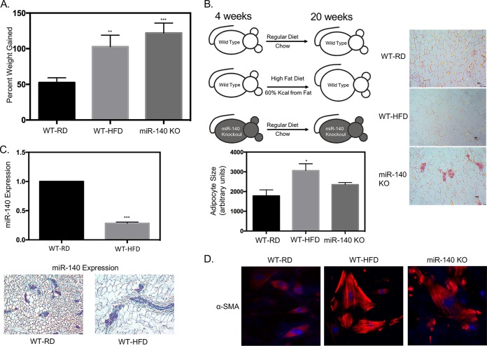
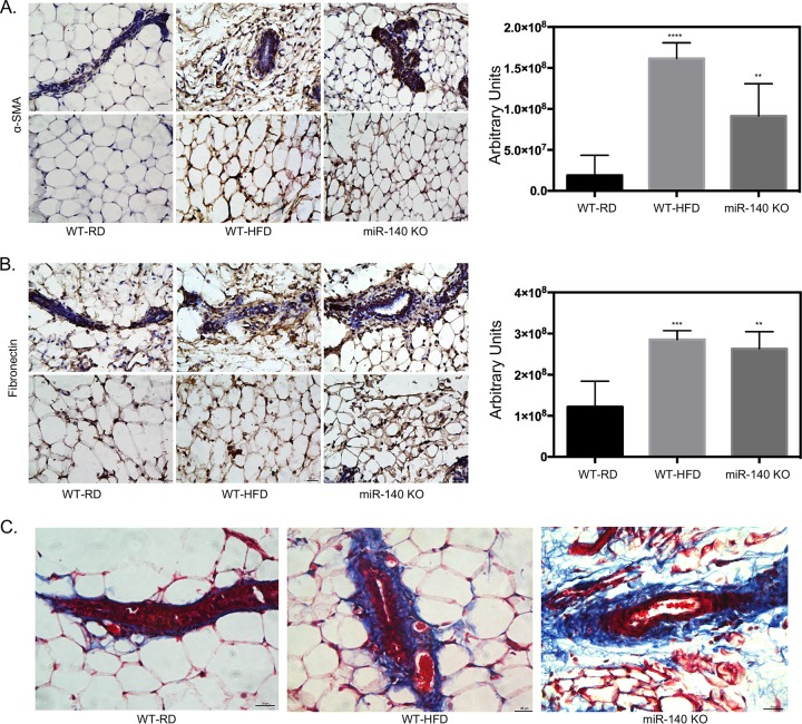
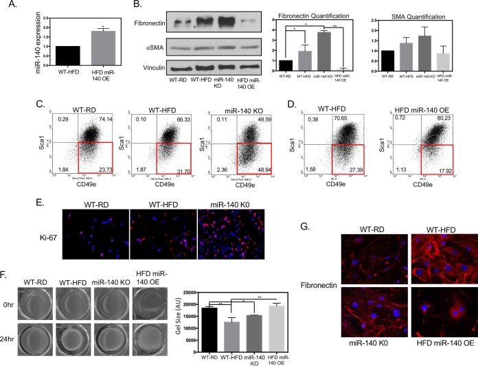
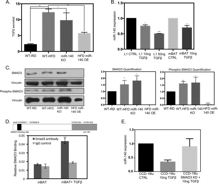
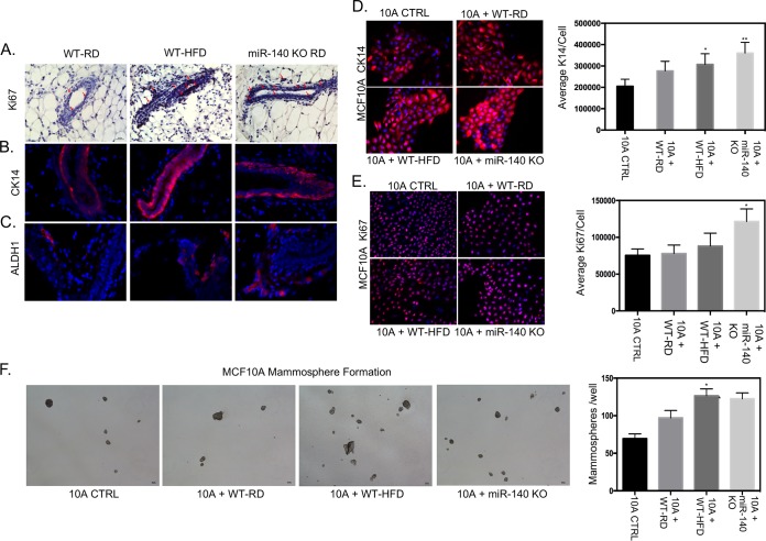
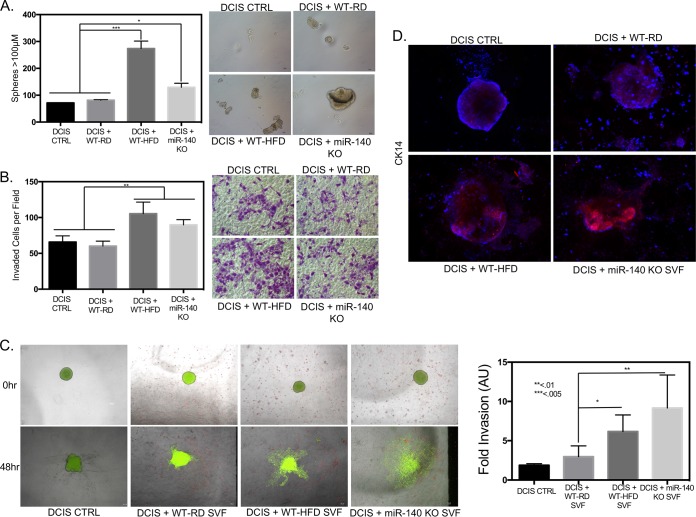
Human breast adipose tissue is a heterogeneous cell population consisting of mature white adipocytes, multipotent mesenchymal stem cells, committed progenitor cells, fibroblasts, endothelial cells, and immune cells. Dependent on external stimulation, adipose-derived stem cells differentiate along diverse lineages into adipocytes, chondrocytes, osteoblasts, fibroblasts, and myofibroblasts. It is currently not fully understood how a high-fat diet reprograms adipose-derived stem cells into myofibroblasts. In our study, we used mouse models of a regular diet and of high-fat-diet-induced obesity to investigate the role of dietary fat on myofibroblast differentiation in the mammary stromal microenvironment. We found that a high-fat diet promotes myofibroblast differentiation by decreasing microRNA 140 (miR-140) expression in mammary adipose tissue through a novel negative-feedback loop. Increased transforming growth factor β1 (TGF-β1) in mammary adipose tissue in obese mice activates SMAD3 signaling, causing phospho-SMAD3 to bind to the miR-140 locus and inhibit miR-140 transcription. This prevents miR-140 from targeting SMAD3 for degradation, resulting in amplified TGF-β1/SMAD3 signaling and miR-140 downregulation-dependent myofibroblast differentiation. Using tissue and coculture models, we found that myofibroblasts and the fibrotic microenvironment created by myofibroblasts impact the stemness and proliferation of normal ductal epithelial cells and early-stage breast cancer invasion and stemness.

Keywords: Obesity; breast cancer; fibrosis; miRNA; myofibroblasts.


Figures

FIG 1
FIG 1
High-fat diet downregulates miR-140 in mammary stromal cells. Female wild-type C57BL/6 mice were fed a regular chow diet (WT-RD) or a high-fat diet (WT-HFD) ad libitum for 16 weeks. (A) Percent weight gained after 16 weeks of a regular or high-fat diet. (B) Left panel, WT-RD, WT-HFD, and miR-140 KO mice after the respective diets for 16 weeks (left panel). Right panels, hematoxylin-and-eosin (H&E) staining of stromal areas of the mammary fat pads isolated from WT-RD, WT-HFD, and miR-140 KO mice. Bottom panel, quantification of adipocyte size. (C) miR-140 is downregulated in mammary stromal cells from high-fat-diet mice. Top panel, miR-140 expression was measured using qRT-PCR of RNA isolated from mammary fat pad SVF cells from WT-RD and obese mice. Bottom panels, RNA in situ staining for miR-140 expression in the mammary fat pads of WT-RD and WT-HFD mice verified downregulation in stromal cells. (D) Immunofluorescence detection of myofibroblast marker αSMA showed upregulation in SVF cells isolated from the mammary fat pads of obese and miR-140 KO mice 10 days after adipogenesis was induced in vitro. *, P < 0.05; **, P < 0.005; ***, P < 0.0001.
FIG 2
FIG 2
High-fat diet-induced miR-140 downregulation is associated with increased ECM deposition. Staining for markers of fibrosis in mammary fat pad tissue sections isolated from WT-RD, WT-HFD, and miR-140 KO mice is shown. (A) Immunohistochemistry staining for αSMA shows increased αSMA expression in the stromal cells and surrounding the mammary ducts of WT-HFD and miR-140 KO mouse mammary fat pads. (B) Immunohistochemistry staining for fibronectin shows increased fibronectin expression in the stromal cells and surrounding the mammary ducts of WT-HFD and miR-140 KO mouse mammary adipose tissue. (C) Modified Masson's trichrome stain shows increased collagen deposition (blue) in WT-HFD and miR-140 KO mouse mammary adipose tissue. **, P < 0.005; ***, P < 0.0005; ****, P < 0.0001.
FIG 3
FIG 3
miR-140 deficiency contributes to myofibroblast differentiation. Protein, cellular, and functional markers of myofibroblast differentiation were examined in SVF cells isolated from the mammary fat pads of WT-RD, WT-HFD, and miR-140 KO mice. (A) Transient transfection was performed to rescue miR-140 expression in WT-HFD mouse SVF cells. (B) Expression levels of proteins associated with fibrosis and myofibroblast differentiation were analyzed using Western blotting. SVF cells from obese and miR-140 KO mice had higher expression levels of fibronectin and αSMA compared to WT-RD. Transient overexpression (OE) of miR-140 in SVF cells from obese mice resulted in decreased expression of all markers examined. (C) Myofibroblast formation was assessed using Sca-1 and CD49e markers using FACS analysis. SVF cells from obese and miR-140 KO mice demonstrated increased myofibroblast populations (SCA-1low CD49ehigh). (D) FACS sorting for myofibroblasts in WT-HFD mouse SVF and WT-HFD mouse SVF overexpressing miR-140. (E) Ki-67 expression was examined in mammary fat pad tissue sections. Ki-67 was found to be upregulated in WT-HFD and miR-140 KO mouse mammary adipose tissue. (F) Evaluation of myofibroblast differentiation using a collagen gel contraction assay. Cells from obese and miR-140 KO mice exhibited higher contractile ability than those from WT-RD mice. Overexpression of miR-140 almost completely ablated contractile ability. The contraction capacity was calculated by measuring the gel area with ImageJ 24 h after the stress was lifted. (G) The amount of fibronectin secreted by stromal vascular fraction cells was examined by immunofluorescence. Data are means ± standard deviations (SD) (n = 3). *, P < 0.05; **, P < 0.005.
FIG 4
FIG 4
TGF-β1 signaling and miR-140 form a negative-feedback loop. TGF-β1 downregulates miR-140 through SMAD3 binding to the miR-140 upstream sequence. (A) High levels of TGF-β1 are secreted by SVF cells from obese and miR-140 KO mice. (B) TGF-β1 decreases miR-140 expression in a dose-dependent manner in 3T3-L1 and mBAT mouse preadipocytes. (C) SMAD3 and phospho-SMAD3 protein expression is upregulated in SVF cells from obese and miR-140 KO mice, indicating increased pathway activity. miR-140 overexpression (OE) in WT-HFD mouse cells completely downregulated SMAD3/pSMAD3 expression. (D) Putative SMAD3 binding site in the upstream sequence of miR-140. ChIP demonstrates increased SMAD3 binding to miR-140 when mBAT cells are treated with 10 ng/ml of TGF-β1. (E) SMAD3 knockdown prevented TGF-β1-mediated miR-140 downregulation in CCD-19lu human lung fibroblasts. *, P < 0.05; **, P < 0.01.
FIG 5
FIG 5
Effects of a high-fat diet on mammary epithelial cells. To investigate the effect of mammary fat pad fibrosis and high-fat diet on mammary epithelial cells, markers of proliferation, basal cell lineage, and stemness were examined in ductal epithelial tissue sections isolated from WT-RD, WT-HFD, and miR-140 KO mice. The immortalized epithelial cell line MCF10A was examined for the same markers after being treated with control SVF medium (10A CTRL) or conditioned medium from SVF cell lines. (A) Immunohistochemistry staining for Ki-67 demonstrated an increase in ductal epithelial cells from obese and miR-140 KO mice. (B) Immunofluorescence staining for basal cell lineage marker CK14 found increased expression in ductal epithelial cells from obese and miR-140 KO mice. (C) Immunofluorescence staining showed that stem cell marker ALDH1 was upregulated in ductal epithelial cells from obese and miR-140 KO mice. (D and E) MCF10A cells were treated with conditioned medium from SVF cells for 48 h before immunofluorescence staining. (D) MCF10A cells treated with conditioned medium from obese and miR-140 KO mouse SVF cells had significantly increased CK14 expression. (E) MCF10A cells treated with conditioned medium from obese-mouse SVF cells had a small increase in Ki-67 expression. MCF10A cells treated with conditioned medium from SVF cells from miR-140 KO mice had significantly increased Ki-67 expression. (F) MCF10A cells treated with conditioned medium from SVF cells from obese and miR-140 KO mice demonstrated significantly increased mammosphere formation. Data are means ± SD (n = 3). *, P < 0.05; **, P < 0.005.
FIG 6
FIG 6
Mammary stromal cells from high-fat-diet and miR-140 KO mice promote breast cancer. DCIS cells were cocultured with SVF cells and then replated for functional assays to examine the effect of fibrotic SVF cells on DCIS invasiveness and aggression. (A) DCIS cells were cocultured with SVF cells for 48 h and then replated for mammosphere formation. DCIS cells cocultured with SVF cells from obese and miR-140 KO mice had significantly increased mammosphere formation. (B) After coculture, DCIS cells were plated for a transwell Matrigel invasion assay. (C) DCIS organoids were plated in a 3D Matrigel culture with SVF cells. After 48 h, organoid invasion was measured using ImageJ. DCIS organoids in 3D culture with SVF cells from obese and miR-140 KO mice had much higher levels of 3D invasion than DCIS cells cultured with SVF cells from WT-RD mice or alone. (D) At 48 h after organoid implantation, DCIS organoids in the 3D Matrigel culture with SVF cells were stained for a marker of basal lineage and tumor invasion/aggression, CK14. DCIS organoids cultured with SVF cells from obese and miR-140 KO mice had increased CK14 expression. Data are means ± SD (n = 3). DCIS CTRL, DCIS cells without coculture. *, P < 0.01; **, P < 0.005; ***, P < 0.001.

References

    1. Gimble JM, Guilak F, Nuttall ME, Sathishkumar S, Vidal M, Bunnell BA. 2008. In vitro differentiation potential of mesenchymal stem cells. Transfus Med Hemother 35:228–238. doi: 10.1159/000124281. - DOI - PMC - PubMed
    1. Kalluri R, Zeisberg M. 2006. Fibroblasts in cancer. Nat Rev Cancer 6:392–401. doi: 10.1038/nrc1877. - DOI - PubMed
    1. Chandler EM, Seo BR, Califano JP, Andresen Eguiluz RC, Lee JS, Yoon CJ, Tims DT, Wang JX, Cheng L, Mohanan S, Buckley MR, Cohen I, Nikitin AY, Williams RM, Gourdon D, Reinhart-King CA, Fischbach C. 2012. Implanted adipose progenitor cells as physicochemical regulators of breast cancer. Proc Natl Acad Sci U S A 109:9786–9791. doi: 10.1073/pnas.1121160109. - DOI - PMC - PubMed
    1. Kojima Y, Acar A, Eaton EN, Mellody KT, Scheel C, Ben-Porath I, Onder TT, Wang ZC, Richardson AL, Weinberg RA, Orimo A. 2010. Autocrine TGF-beta and stromal cell-derived factor-1 (SDF-1) signaling drives the evolution of tumor-promoting mammary stromal myofibroblasts. Proc Natl Acad Sci U S A 107:20009–20014. doi: 10.1073/pnas.1013805107. - DOI - PMC - PubMed
    1. Wolters PJ, Collard HR, Jones KD. 2014. Pathogenesis of idiopathic pulmonary fibrosis. Annu Rev Pathol 9:157–179. doi: 10.1146/annurev-pathol-012513-104706. - DOI - PMC - PubMed

Publication types

MeSH terms

LinkOut - more resources