. 2017 May;28(5):1491-1505.
doi: 10.1681/ASN.2015080885. Epub 2016 Nov 28.

Sildenafil Prevents Podocyte Injury via PPAR- γ-Mediated TRPC6 Inhibition

Affiliations

Sildenafil Prevents Podocyte Injury via PPAR- γ-Mediated TRPC6 Inhibition

Ramon Sonneveld et al. J Am Soc Nephrol. 2017 May.

Abstract

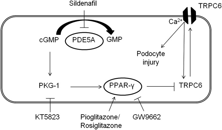
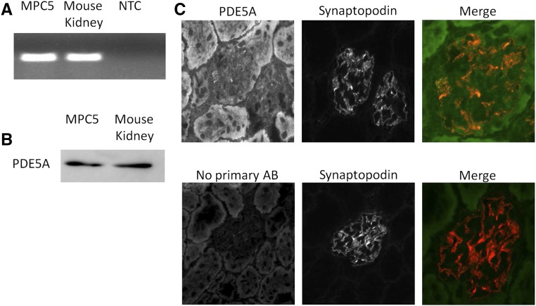
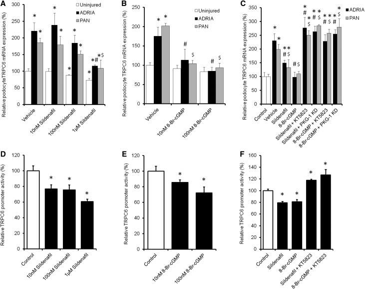
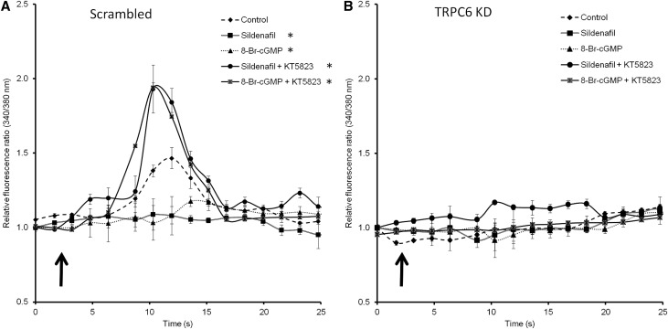
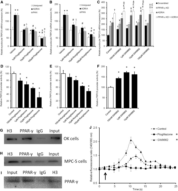
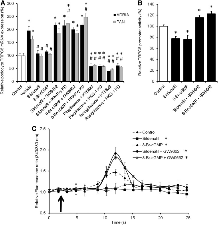
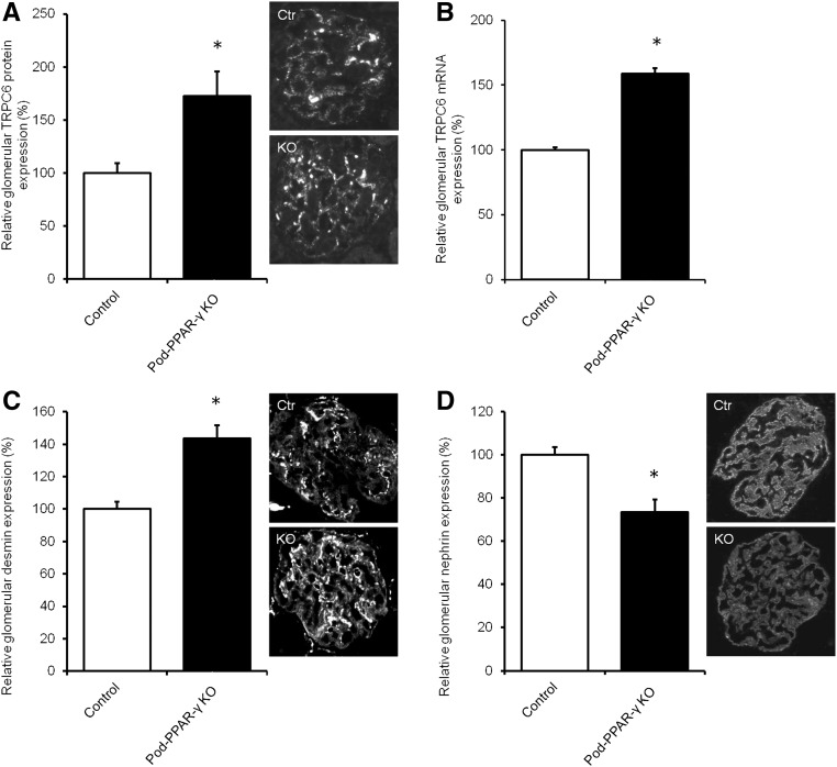
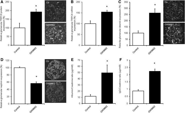
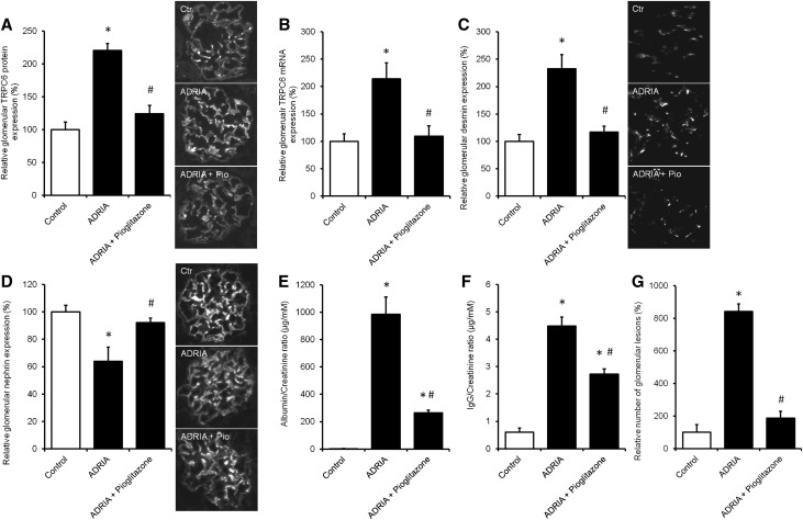
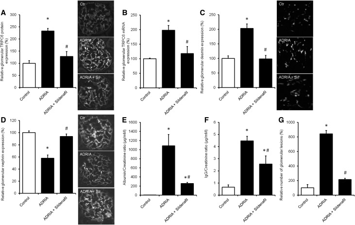
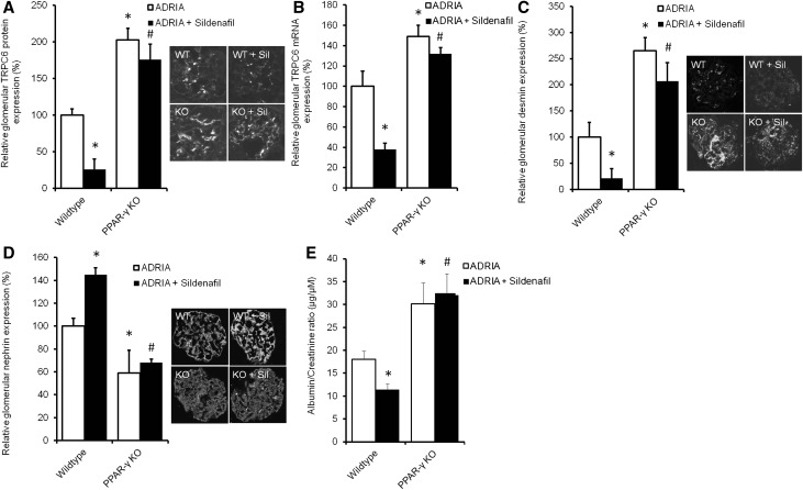
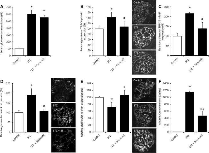
Transient receptor potential channel C6 (TRPC6) gain-of-function mutations and increased TRPC6 expression in podocytes induce glomerular injury and proteinuria. Sildenafil reduces TRPC6 expression and activity in nonrenal cell types, although the mechanism is unknown. Peroxisome proliferator-activated receptor γ (PPAR-γ) is a downstream target of sildenafil in the cyclic guanosine monophosphate (cGMP)-activated protein kinase G (PKG) axis. PPAR-γ agonists, like pioglitazone, appear antiproteinuric. We hypothesized that sildenafil inhibits TRPC6 expression in podocytes through PPAR-γ-dependent mechanisms, thereby counteracting podocyte injury and proteinuria. Treatment with sildenafil, the cGMP derivative 8-bromoguanosine 3',5'-cyclic monophosphate sodium salt (8-Br-cGMP), or pioglitazone dose-dependently downregulated podocyte injury-induced TRPC6 expression in vitro Knockdown or application of antagonists of PKG or PPAR-γ enhanced TRPC6 expression in podocytes and counteracted effects of sildenafil and 8-Br-cGMP. We observed similar effects on TRPC6 promoter activity and TRPC6-dependent calcium influx. Chromatin immunoprecipitation showed PPAR-γ binding to the TRPC6 promoter. Sildenafil or pioglitazone treatment prevented proteinuria and the increased TRPC6 expression in rats with adriamycin-induced nephropathy and mice with hyperglycemia-induced renal injury. Rats receiving PPAR-γ antagonists displayed proteinuria and increased podocyte TRPC6 expression, as did podocyte-specific PPAR-γ knockout mice, which were more sensitive to adriamycin and not protected by sildenafil. Thus, sildenafil ameliorates podocyte injury and prevents proteinuria through cGMP- and PKG-dependent binding of PPAR-γ to the TRPC6 promoter, which inhibits TRPC6 promoter activity, expression, and activity. Because sildenafil is approved for clinical use, our results suggest that additional clinical study of its antiproteinuric effect in glomerular disease is warranted.

Keywords: albuminuria; focal segmental glomerulosclerosis; glomerulosclerosis; ion channel; podocyte; signaling.


Figures

Figure 1.
Figure 1.
Hypothesis that links sildenafil to TRPC6. Sildenafil inhibits PDE5A, thereby indirectly increasing cellular cGMP levels. cGMP signaling activates PKG-1 and subsequently PPAR-γ, which inhibits TRPC6 transcription and expression. This leads to reduced TRPC6 channel expression at the membrane of the podocyte, thereby reducing TRPC6–mediated Ca2+ influx and eventually, podocyte injury in the context of glomerular disease. We propose that this pathway can be modulated by blocking PKG-1 with KT5823, activating PPAR-γ with pioglitazone or rosiglitazone, or inhibiting PPAR-γ with GW9662.
Figure 2.
Figure 2.
PDE5A is expressed by podocytes. RNA and protein were isolated from cultured podocytes and renal mouse cortex; subsequently, (A) PCR and (B) Western blot were performed to determine PDE5A expression in these samples. Glomerular PDE5A expression was confirmed by immunofluorescence staining of PDE5A. (C) Importantly, costaining with synaptopodin showed PDE5A expression in the podocytes; merge is a higher magnification of single-channel images. AB, antibody; NTC, no template control.
Figure 3.
Figure 3.
Sildenafil dose-dependently downregulates TRPC6 expression via PKG-1 in injured podocytes by affecting TRPC6 promoter activity. Cultured podocytes, transfected with either PKG-1 or scrambled siRNA, were injured by adriamycin (ADRIA) or PAN application and treated with different concentrations of (A) sildenafil or (B) 8-Br-cGMP (C) with or without PKG-1 antagonist KT5823, and TRPC6 mRNA expression was determined. Opossum Kidney cells expressing a TRPC6 promoter luciferase construct were treated with the same concentrations of (D) sildenafil or (E) 8-Br-cGMP (F) with or without KT5823. Firefly-to-Renilla ratio was calculated and normalized for vehicle-treated cells; n=4–8 in at least two independent experiments. Statistical significance was determined using ANOVA followed by Bonferroni post hoc test. *P<0.05 versus control cells; #P<0.05 versus ADRIA-treated cells; $P<0.05 versus PAN-treated cells.
Figure 4.
Figure 4.
Downregulation of TRPC6 expression by sildenafil leads to reduced TRPC6–mediated Ca2+ influx into the podocyte. Intracellular Ca2+ concentration was determined by fura-2 ratiometry. Cultured podocytes transfected with a scrambled siRNA construct were pretreated with sildenafil or 8-Br-cGMP with or without the PKG-1 antagonist KT5823 for 24 hours. (A) After removal of the specific media, cells were exposed to 100 μM 1-oleoyl-2-acetyl-sn-glycerolin (OAG) to activate the TRPC6 ion channel. (B) The same experimental setup was tested in TRPC6 KD podocytes. The arrows indicate the time points of OAG application; n=5 in two independent experiments. Statistical significance was determined using a repeated measurement test. *P<0.05 versus vehicle-treated cells.
Figure 5.
Figure 5.
PPAR-γ agonists and antagonists as well as PPAR-γ KD influence TRPC6 promoter activity, TRPC6 expression, and channel activity. Cultured podocytes were injured by adriamycin (ADRIA) or PAN application and treated with different concentrations of the PPAR-γ agonist (A) pioglitazone or (B) rosiglitazone, and TRPC6 mRNA expression was determined. (C) In addition, TRPC6 expression was determined in uninjured podocytes, transfected with scrambled or PPAR-γ siRNA, and treated with various concentrations of the PPAR-γ antagonist GW9662. A luciferase assay was performed to determine TRPC6 promoter activity in Opossum Kidney cells treated with (D) pioglitazone, (E) rosiglitazone, and (F) GW9662. TRPC6-to-GAPDH and Firefly-to-Renilla ratios were calculated and normalized for vehicle-treated cells. ChIP assays were performed to determine whether PPAR-γ directly binds to the TRPC6 promoter. This was tested using (G) a promoter construct transfected into OK cells or (H) the endogenous TRPC6 promoter in cultured mouse podocytes. Antibodies against histone H3 (H3) as positive control, PPAR-γ to determine whether PPAR-γ bound to the TRPC6 promoter, and IgG as negative control were used to purify fractions of the DNA. (G and H) Hereafter, the proteins were digested, PCR was performed with primers detecting the TRPC6 promoter, and products were put on gel. (I) Western blot analysis showed that PPAR-γ was present in the input sample, in the PPAR-γ–immunoprecipitated sample, and in the H3-positive control sample of the nontransfected MPC-5 cells. Cultured podocytes stably transfected with scrambled siRNA were pretreated with sildenafil or 8-Br-cGMP with or without KT5823 for 24 hours. (J) After removal of the specific media, cells were exposed to 100 μM 1-oleoyl-2-acetyl-sn-glycerolin (OAG) to activate the TRPC6 ion channel. Intracellular Ca2+ concentration was determined by fura-2 ratiometry. The arrow indicates OAG application; n=4–8, in at least two independent experiments. Statistical significance was determined using ANOVA followed by (A–F) Bonferroni post hoc test or (J) repeated measurement test. *P<0.05 versus vehicle-treated uninjured cells; #P<0.05 versus ADRIA-treated cells; $P<0.05 versus PAN-treated cells; P<0.05 versus scrambled transfected uninjured cells; ¥P<0.05 versus the GW9662 of equal concentration-treated scrambled cells; £P<0.05 versus the GW9662 of equal concentration-treated PPAR-γ KD cells; ±P<0.05 versus the ADRIA-challenged GW9662 of equal concentration-treated cells.
Figure 6.
Figure 6.
PPAR-γ antagonism abrogates the effect of sildenafil on TRPC6 promoter activity, TRPC6 expression, and channel activity. Cultured podocytes, transfected with scrambled, PKG-1, or PPAR-γ siRNA, were injured by adriamycin (ADRIA) or PAN application and treated with sildenafil, 8-Br-cGMP, pioglitazone, or rosiglitazone with or without PPAR-γ antagonist GW9662. (A) Subsequently, TRPC6 mRNA expression was determined. (B) TRPC6 promoter activity was measured using a luciferase promoter assay in Opossum Kidney cells treated with sildenafil or 8-Br-cGMP with or without GW9662. Firefly-to-Renilla ratio was calculated and normalized for vehicle-treated cells. Cultured podocytes stably transfected with scrambled siRNA were pretreated with sildenafil or 8-Br-cGMP with or without GW9662 for 24 hours. (C) After removal of the specific media, cells were exposed to 100 μM 1-oleoyl-2-acetyl-sn-glycerolin (OAG) to activate the TRPC6 ion channel. Intracellular Ca2+ concentration was determined by fura-2 ratiometry. The arrow indicates the time point of OAG application; n=4–7 in at least two independent experiments. Statistical significance was determined using ANOVA followed by (A and B) Bonferroni post hoc test or (C) repeated measurement test. *P<0.05 versus vehicle-treated cells; #P<0.05 versus ADRIA-treated cells.
Figure 7.
Figure 7.
Podocyte-specific PPAR-γ–deficient mice show increased glomerular TRPC6 expression. Glomerular TRPC6 (A) protein and (B) mRNA expression was determined. In addition, protein expression of (C) desmin and (D) nephrin was measured in podocyte–specific PPAR-γ–deficient (KO) mice as well as their wild-type littermates. Protein expression is depicted relative to the mean expression in wild-type mice; n=4 animals per group. Statistical significance was determined using an unpaired t test. Ctr, control. *P<0.05 versus wild-type littermates.
Figure 8.
Figure 8.
Pharmacologic inhibition of PPAR-γ in rats increases glomerular TRPC6 expression and leads to podocyte injury and proteinuria. Wistar rats were treated with the PPAR-γ antagonist GW9662 (2.5 mg/kg body wt) for 3 weeks, after which animals were euthanized, and glomerular TRPC6 (A) protein and (B) mRNA expression was determined. In addition, protein expression of (C) desmin and (D) nephrin was measured. Protein expression is depicted as percentage relative to the mean expression in vehicle-treated rats. In addition, (E) urinary albumin-to-creatinine ratio (micrograms per millimolar) and (F) IgG-to-creatinine ratio (micrograms per millimolar) were assessed; n=4 animals per group. Statistical significance was determined using an unpaired t test. Ctr, control. *P<0.05 versus control rats.
Figure 9.
Figure 9.
Pioglitazone treatment prevents TRPC6 overexpression and podocyte injury in the adriamycin (ADRIA)–induced nephropathy rat model for FSGS. ADRIA-induced nephropathy was induced in rats, after which control and ADRIA nephropathy rats were treated with the PPAR-γ agonist pioglitazone (12 mg/kg body wt) for 6 weeks. Thereafter, animals were euthanized, and glomerular TRPC6 (A) protein and (B) mRNA expression was determined. In addition, protein expression of (C) desmin and (D) nephrin was measured. Protein expression is depicted as percentage of vehicle-treated rats. In addition, (E) urinary albumin-to-creatinine ratio (micrograms per millimolar), (F) IgG-to-creatinine ratio (micrograms per millimolar), and (G) percentage of glomerular lesions were assessed; n=8 animals per group. Statistical significance was determined using ANOVA followed by Bonferroni post hoc test. Ctr, control. *P<0.05 versus vehicle-treated control rats; #P<0.05 versus ADRIA–treated control rats.
Figure 10.
Figure 10.
Sildenafil treatment prevents TRPC6 overexpression and podocyte injury in the adriamycin (ADRIA)–induced nephropathy rat model for FSGS. ADRIA-induced nephropathy was induced in rats, after which control and ADRIA nephropathy rats were treated with sildenafil (5 mg/kg body wt) for 6 weeks. Thereafter, animals were euthanized, and glomerular TRPC6 (A) protein and (B) mRNA expression was determined. In addition, protein expression of (C) desmin and (D) nephrin was measured. Protein expression is depicted as percentage of vehicle-treated rats. In addition, (E) urinary albumin-to-creatinine ratio (micrograms per millimolar), (F) IgG-to-creatinine ratio (micrograms per millimolar), and (G) percentage of glomerular lesions were assessed; n=8 animals per group. Statistical significance was determined using ANOVA followed by Bonferroni post hoc test. Ctr, control. *P<0.05 versus vehicle-injected control rats; #P<0.05 versus ADRIA–treated control rats.
Figure 11.
Figure 11.
Sildenafil does not prevent TRPC6 overexpression and podocyte injury in adriamycin (ADRIA)–challenged podocyte-specific PPAR-γ KO mice. ADRIA-induced nephropathy was induced in podocyte-specific PPAR-γ KO and wild-type mice, after which they were treated with sildenafil. Thereafter, animals were euthanized, and glomerular TRPC6 (A) protein and (B) mRNA expression was determined. In addition, protein expression of (C) desmin and (D) nephrin was measured. Protein expression is depicted as percentage of vehicle-treated mice. (E) In addition, urinary albumin-to-creatinine ratio (micrograms per micromolar) was assessed; n=3–8 animals per group. Statistical significance was determined using ANOVA followed by Bonferroni post hoc test. WT, wild type.*P<0.05 versus vehicle-injected control mice; #P<0.05 versus ADRIA–treated control mice.
Figure 12.
Figure 12.
Sildenafil treatment prevents TRPC6 overexpression and podocyte injury in the STZ–induced hyperglycemic mouse model for renal injury. STZ-induced hyperglycemia was induced in healthy mice (STZ), after which animals were treated with sildenafil. Thereafter, animals were euthanized, and (A) serum glucose and glomerular TRPC6 (B) protein and (C) mRNA expression were determined. In addition, protein expression of (D) desmin and (E) nephrin was measured, and (F) urinary albumin-to-creatinine ratio (micrograms per milligram) was assessed. Protein expression is depicted as percentage of vehicle-treated mice; n=3–5 animals per group. Statistical significance was determined using ANOVA followed by Bonferroni post hoc test. *P<0.05 versus vehicle-injected control mice; #P<0.05 versus STZ-treated mice.

Comment in

  • The Use of Sildenafil for Glomerular Disease.
    Tardi NJ, Reiser J. Tardi NJ, et al. J Am Soc Nephrol. 2017 May;28(5):1329-1331. doi: 10.1681/ASN.2017020171. Epub 2017 Mar 30. J Am Soc Nephrol. 2017. PMID: 28360220 Free PMC article. No abstract available.

References

    1. Ballard SA, Gingell CJ, Tang K, Turner LA, Price ME, Naylor AM: Effects of sildenafil on the relaxation of human corpus cavernosum tissue in vitro and on the activities of cyclic nucleotide phosphodiesterase isozymes. J Urol 159: 2164–2171, 1998 - PubMed
    1. Shulman AI, Mangelsdorf DJ: Retinoid x receptor heterodimers in the metabolic syndrome. N Engl J Med 353: 604–615, 2005 - PubMed
    1. Liu HF, Guo LQ, Huang YY, Chen K, Tao JL, Li SM, Chen XW: Thiazolidinedione attenuate proteinuria and glomerulosclerosis in Adriamycin-induced nephropathy rats via slit diaphragm protection. Nephrology (Carlton) 15: 75–83, 2010 - PubMed
    1. Yang HC, Ma LJ, Ma J, Fogo AB: Peroxisome proliferator-activated receptor-gamma agonist is protective in podocyte injury-associated sclerosis. Kidney Int 69: 1756–1764, 2006 - PubMed
    1. Jung JH, Kim BJ, Chae MR, Kam SC, Jeon JH, So I, Chung KH, Lee SW: Gene transfer of TRPC6 (dominant negative) restores erectile function in diabetic rats. J Sex Med 7: 1126–1138, 2010 - PubMed