. 2017 Feb;91(2):100-109.
doi: 10.1124/mol.116.106484. Epub 2016 Nov 28.

The E Loop of the Transmitter Binding Site Is a Key Determinant of the Modulatory Effects of Physostigmine on Neuronal Nicotinic α4β2 Receptors

Affiliations

The E Loop of the Transmitter Binding Site Is a Key Determinant of the Modulatory Effects of Physostigmine on Neuronal Nicotinic α4β2 Receptors

Xiaochun Jin et al. Mol Pharmacol. 2017 Feb.

Abstract

Physostigmine is a well known inhibitor of acetylcholinesterase, which can also activate, potentiate, and inhibit acetylcholine receptors, including neuronal nicotinic receptors comprising α4 and β2 subunits. We have found that the two stoichiometric forms of this receptor differ in the effects of physostigmine. The form containing three copies of α4 and two of β2 was potentiated at low concentrations of acetylcholine chloride (ACh) and physostigmine, whereas the form containing two copies of α4 and three of β2 was inhibited. Chimeric constructs of subunits indicated that the presence of inhibition or potentiation depended on the source of the extracellular ligand binding domain of the subunit. Further sets of chimeric constructs demonstrated that a portion of the ACh binding domain, the E loop, is a key determinant. Transferring the E loop from the β2 subunit to the α4 subunit resulted in strong inhibition, whereas the reciprocal transfer reduced inhibition. To control the number and position of the incorporated chimeric subunits, we expressed chimeric constructs with subunit dimers. Surprisingly, incorporation of a subunit with an altered E loop had similar effects whether it contributed either to an intersubunit interface containing a canonical ACh binding site or to an alternative interface. The observation that the α4 E loop is involved suggests that physostigmine interacts with regions of subunits that contribute to the ACh binding site, whereas the lack of interface specificity indicates that interaction with a particular ACh binding site is not the critical factor.


Figures

Fig. 1.
Fig. 1.
Effect of transplanting the E loop between the α4 and β2 subunits. Traces are shown from oocytes when ACh was initially applied alone and then the perfusion was switched to ACh + physostigmine (ACh alone application indicated by the lower line above the trace, and the time of the application of both ACh + physostigmine shown by the upper line). The constructs injected are shown above the trace (constructs are indicated with & separating the independent constructs, e.g. α&β 8:1 indicates that free subunits were injected at a ratio of 8:1::α4:β2, while α-β&α indicates that the α4-β2 dimer was injected with free α4 subunit); Panel A: α4&β2 8:1; B α4&β2 1:8; C α4-β2 & β2; D β2-α4 & β2; E α4-β2 & β2(E); F β2-α4 & β2(E); G α4-β2 & α4; H β2-α4 & α4; I α4-β2 & α4(E); J β2-α4 & α4(E). The horizontal bar shows 20 sec for all traces while the vertical bar shows the current calibration for each trace. The ACh concentration was adjusted to result in a response of less than 20% of the maximal response for that oocyte. Concentrations used were: A 1 µM ACh; B 0.3 µM; C 0.3 µM; D 0.3 µM; E 0.3 µM; F 0.1 µM; G 1 µM; H 1 µM; I 0.3 µM; J 0.3 µM. 15 µM physostigmine was used for all traces.
Fig. 2.
Fig. 2.
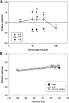
Concentration and voltage dependence of physostigmine actions. (A) The effect of different concentrations of physostigmine on responses from oocytes injected with free α4 and β2 subunits at the indicated ratios (means ± S.E.M.). Data are from 6–40 oocytes, except for the point at 10 µM physostigmine on α4&β2 1:8, which is a single experiment. The P value symbols indicate that the value differs from 1 (no effect) by chance [one-sample t test; ns indicates P > 0.05 (not significant); **P < 0.01; ***P < 0.001]. (B) The inhibition of responses from oocytes injected with α4&β2 1:8 (0.3 µM ACh, 15 µM physostigmine) at different voltages. Symbols show means ± S.E.M. for four oocytes tested at both −50 and −100 mV (“cells at two voltages”) and for 10 eggs tested at −50 mV (“mean” including the four tested at both voltages). The heavy line shows the average relative response for four oocytes subjected to a voltage ramp (see the Materials and Methods), whereas the dashed lines show the standard error.
Fig. 3.
Fig. 3.
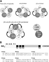
Cartoons of receptor structure. (A) The arrangement of subunits in the receptors formed after injections of various combinations of subunits. The diagrams are of the receptor viewed from the extracellular side. Stars indicate the locations of canonical ACh binding sites (α4/β2 interface). The contributions of loops to a canonical ACh binding site are also shown. (B) A linear diagram of a generic nicotinic subunit with the relative positions of the binding site loops (A–F) and the TM domains (TM1–TM4) is shown. Extracellular portions are indicated by white shading, intracellular by gray shading and TM regions by black. The residues transferred in the chimeric constructs, together with the initial residue number in the mature subunit, are shown below the diagram.
Fig. 4.
Fig. 4.
Location of residues photolabeled by physostigmine or implicated in the actions of Zn2+. (A) Sections of aligned sequence for segments of the extracellular domains of the human α4 and β2 subunits and the Torpedo α1, γ, and δ subunits. The letters on a blue background above the sequences indicate the locations of chimera segments at the (+) side of an interface (loops A and C), whereas those on the orange background indicate those on the (−) side (E and F). Residues are color coded to indicate that they were labeled by physostigmine only in the absence of carbamylcholine (green, in the α1 subunit) or were labeled by physostigmine in the presence of carbamylcholine (red, in γ and δ subunits). Note that no label was reported from the β1 subunit, and it was not possible to sequence the E loop of the δ subunit. Yellow backgrounds indicate residues implicated in the actions of Zn2+: α4H(165) participates in potentiation at the α4/α4 interface and inhibition at the β2/α4 interface, whereas α4(E194) participates in potentiation and β2(D193) in inhibition. The three residues in the E loops of α4 and β2 highlighted in gray are the residues switched in the studies by Harpsøe et al. (2011) referred to in the Discussion. (B) A homology model of the extracellular domains of two α4 subunits, based on the GluCl structure (3RIF; Hibbs and Gouaux, 2011) to provide an idea of the positions of the residues highlighted in (A). The view is from the outside of the receptor looking into the binding site for ACh, and the extracellular portion is at the top. The subunit contributing the (+) side is shown as a cyan ribbon. The C loop on the (+) side is colored blue, and the residue in α4 contributing to the Zn2+ potentiating site is shown as yellow spheres. The residues in α1 that were photolabeled by physostigmine are not shown. The subunit contributing the (−) side is shown as a light gray ribbon and the E loop is colored orange. The histidine contributing to both of the Zn2+ sites is shown as yellow spheres. Red spheres indicate the residues homologous to the residues photolabeled by physostigmine and for which carbamylcholine did not protect against labeling. Finally, the residues in the E loop interchanged by Harpsøe et al. (2011) are shown as black spheres. One residue was both interchanged and photolabeled; it is colored purple. Note that the residues contributing to the Zn2+ sites are relatively far from the E loop, whereas several residues photolabeled by physostigmine cluster near the E loop and to residues shown to be important in determining the properties of the receptor.

References

    1. Carbone AL, Moroni M, Groot-Kormelink PJ, Bermudez I. (2009) Pentameric concatenated (alpha4)(2)(beta2)(3) and (alpha4)(3)(beta2)(2) nicotinic acetylcholine receptors: subunit arrangement determines functional expression. Br J Pharmacol 156:970–981. - PMC - PubMed
    1. Colquhoun D. (1998) Binding, gating, affinity and efficacy: the interpretation of structure-activity relationships for agonists and of the effects of mutating receptors. Br J Pharmacol 125:924–947. - PMC - PubMed
    1. Cooper JC, Gutbrod O, Witzemann V, Methfessel C. (1996) Pharmacology of the nicotinic acetylcholine receptor from fetal rat muscle expressed in Xenopus oocytes. Eur J Pharmacol 309:287–298. - PubMed
    1. Dani JA, Bertrand D. (2007) Nicotinic acetylcholine receptors and nicotinic cholinergic mechanisms of the central nervous system. Annu Rev Pharmacol Toxicol 47:699–729. - PubMed
    1. Gotti C, Moretti M, Gaimarri A, Zanardi A, Clementi F, Zoli M. (2007) Heterogeneity and complexity of native brain nicotinic receptors. Biochem Pharmacol 74:1102–1111. - PubMed

LinkOut - more resources