. 2016 Nov 15:7:542.
doi: 10.3389/fphys.2016.00542. eCollection 2016.

Reciprocal Modulation of IK1-INa Extends Excitability in Cardiac Ventricular Cells

Affiliations

Reciprocal Modulation of IK1-INa Extends Excitability in Cardiac Ventricular Cells

Anthony Varghese. Front Physiol. .

Abstract

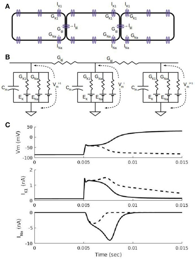
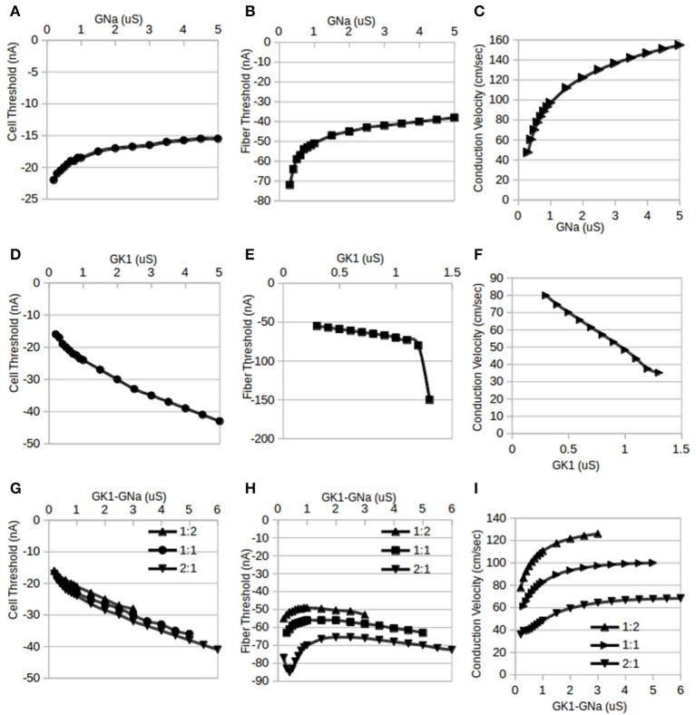
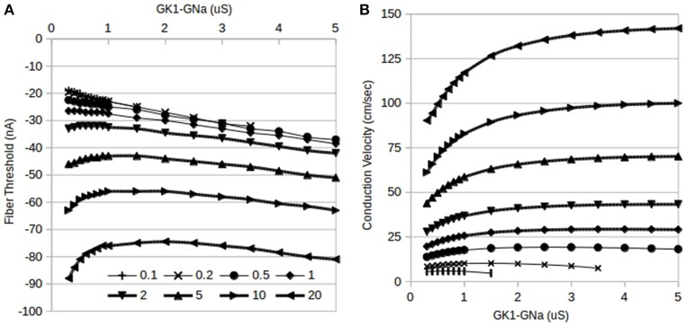
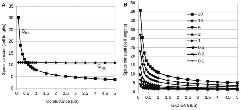
The inwardly rectifying potassium current (IK1) and the fast inward sodium current (INa) are reciprocally modulated in mammalian ventricular myocytes. An increase in the expression of channels responsible for one of these two currents results in a corresponding increase in expression of the other. These currents are critical in the propagation of action potentials (AP) during the normal functioning of the heart. This study identifies a physiological role for IK1-INa reciprocal modulation in ventricular fiber activation thresholds and conduction. Simulations of action potentials in single cells and propagating APs in cardiac fibers were carried out using an existing model of electrical activity in cardiac ventricular myocytes. The conductances, GK1, of the inwardly rectifying potassium current, and GNa, of the fast inward sodium current were modified independently and in tandem to simulate reciprocal modulation. In single cells, independent modulation of GK1 alone resulted in changes in activation thresholds that were qualitatively similar to those for reciprocal GK1-GNa modulation and unlike those due to independent modulation of GNa alone, indicating that GK1 determines the cellular activation threshold. On the other hand, the variations in conduction velocity in cardiac cell fibers were similar for independent GNa modulation and for tandem changes in GK1-GNa, suggesting that GNa is primarily responsible for setting tissue AP conduction velocity. Conduction velocity dependence on GK1-GNa is significantly affected by the intercellular gap junction conductance. While the effects on the passive fiber space constant due to changes in both GK1 and the intercellular gap junction conductance, Ggj, were in line with linear cable theory predictions, both conductances had surprisingly large effects on fiber activation thresholds. Independent modulation of GK1 rendered cardiac fibers inexcitable at higher levels of GK1 whereas tandem GK1-GNa changes allowed fibers to remain excitable at high GK1 values. Reciprocal modulation of the inwardly rectifying potassium current and the fast inward sodium current may have a functional role in allowing cardiac tissue to remain excitable when IK1 is upregulated.

Keywords: cardiac cells; mathematical model; reciprocal modulation.


Figures

Figure 1
Figure 1
Action potential propagation in a cardiac fiber. (A) Schematic view of three cells in a fiber. In addition to a number of sodium, potassium, and calcium currents in the cell membrane, each cell has non-selective gap junction channels that allow currents to flow down the fiber. The gap junction current, Igj, allows a depolarized cell to drive electrical activity in a neighboring cell. (B). Equivalent circuit representation of the three cells in (A). Each cell has a capacitance associated with the cell membrane and ionic currents represented by a conductance and a battery for the Nernst potential for the corresponding ion. The inward rectifier potassium current, IK1, current has a non-linear conductance (see Methods) with a maximal conductance, GK1, and its reversal potential is the potassium Nernst potential, EK. The fast sodium current, INa, has a time-dependent conductance with the maximal conductance, GNa, and its reversal potential is the sodium Nernst potential, ENa. (C) Simulations showing cell membrane potential, Vm, and ion channel currents, IK1 and INa, for a subthreshold stimulus (dashed curves) and suprathreshold stimulus (solid curves). Results shown are for the 5th cell in a fiber of 100 cells with 0.2 ms-duration current stimuli administered at the 5 ms mark to the first 4 cells. A subthreshold stimulus brings Vm to about −60 mV after which Vm declines (dashed curve) back to its resting membrane potential of about −90 mV; the IK1 waveform due to a subthreshold stimulus is larger than the suprathreshold case (solid curve) because the subthreshold Vms are more negative and thus less inward rectified. INa is partially activated (dashed curve) for a subthreshold stimulus and fully activated (solid curve) for the suprathreshold stimulus; the fully activated INa is what triggers the full depolarization of Vm to about +20 Mv.
Figure 2
Figure 2
Independent and reciprocal modulation of IK1 and INa in single cells and fibers. By standard convention, inward (depolarizing) stimulus currents are negative. (A). Changes in single cell threshold due to independent modulation of GNa. These results indicate that as GNa is increased, the magnitude of the threshold is lowered. GK1 was held at its default value of 0.5 μS. (B) Fiber threshold was also lowered as GNa was increased. GK1 was held at 0.5 μS and Ggj was held at 10 μS. (C) Fiber conduction velocity increased as GNa was increased (same conditions as B). (D) Changes in single cell threshold due to independent modulation of GK1: as GK1 is increased, the magnitude of the threshold is raised. GNa was held at 0.5 μS. (E) Fiber threshold was raised as GK1 was increased. For values of GK1 larger than 1.3 μS, the fiber was inexcitable. GNa was held at its default value of 0.5 μS and Ggj was held at 10 μS. (F) Fiber conduction velocity decreased as GK1 was increased (same conditions as D). (G) Changes in single cell threshold due to reciprocal modulation of GK1 and GNa. Cell threshold increased monotonically with the tandem changes in GK1:GNa. Three different ratios for GK1:GNa were used: 1:2, 1:1, and 2:1; the X-axis shows the GK1 values, the corresponding GNa value depends on the ratio used. (H) Fiber threshold changes due to reciprocal modulation of GK1 and GNa. Three different ratios for GK1:GNa were used (1:2, 1:1, and 2:1) while Ggj was held at 10 μS. The X-axis shows the GK1 values, the corresponding GNa value depends on the ratio used. (I) Fiber conduction velocity increased with reciprocal modulation of GK1 and GNa (same conditions as D). The X-axis shows the GK1 values, the corresponding GNa value depends on the ratio used.
Figure 3
Figure 3
Effects of reciprocal modulation of IK1 and INa and changes in gap junction conductance, Ggj, on fiber thresholds. (A) Changes in fiber threshold due to reciprocal modulation of GK1–GNa (1:1 ratio). At any particular value of GK1–GNa, increasing Ggj raises the threshold for initiating a propagating action potential. The curves for Ggj = 0.1 and 0.2 μS (top two curves) overlap considerably. Magnitudes of current thresholds increase monotonically with increasing GK1–GNa for Ggj = 0.1–1 μS while higher values of Ggj result in biphasic dependence. (B) Changes in fiber conduction velocity due to reciprocal modulation of GK1–GNa. At any particular value of GK1–GNa, increasing Ggj increases the conduction velocity of propagating action potentials. Markers are the same as panel A: top curve is for Ggj = 20 μS.
Figure 4
Figure 4
Effects of independent and reciprocal modulation of IK1 and INa and changes in gap junction conductance, Ggj, on fiber space constant. (A) Changes in fiber space constant due to independent modulation of GK1 and GNa: the space constant is insensitive to changes in GNa alone (GK1 held at 0.5 μS–flat line) but is very sensitive to alterations in GK1 (GNa held at 0.5 μS–curve). (B) Changes in fiber space constant due to reciprocal modulation of GK1–GNa (1:1 ratio). Each curve is for a different value of Ggj from 0.1 to 20 μS.

Comment in

References

    1. Balse E., Steele D. F., Abriel H., Coulombe A., Fedida D., Hatem S. N. (2012). Dynamic of ion channel expression at the plasma membrane of cardiomyocytes. Physiol. Rev. 92, 1317–1358. 10.1152/physrev.00041.2011 - DOI - PubMed
    1. Barker R. J., Price R. L., Gourdie R. G. (2002). Increased association of ZO-1 with connexin43 during remodeling of cardiac gap junctions. Circ. Res. 90, 317–332. 10.1161/hh0302.104471 - DOI - PubMed
    1. Bu G., Adams H., Berbari E. J., Rubart M. (2009). Uniform action potential repolarization within the sarcolemma of in situ ventricular cardiomyocytes. Biophys. J. 96, 2532–2546. 10.1016/j.bpj.2008.12.3896 - DOI - PMC - PubMed
    1. Christé G. (1999). Localization of K+ channels in the tubules of cardiomyocytes as suggested by the parallel decay of membrane capacitance, IK1 and IKATP during culture and by delayed IK1 response to barium. J. Mol. Cell. Cardiol. 31, 2207–2213. 10.1006/jmcc.1999.1034 - DOI - PubMed
    1. Crawford J. D., Golubitsky M., Gomes M. G. M., Knobloch E., Stewart I. N. (1991). Boundary conditions as symmetry constraints, in Singularity Theory and its Applications: Warwick 1989, Part II: Singularities, Bifurcations and Dynamics, ed Roberts M. (Berlin: Springer; ), 63–79.

LinkOut - more resources