

Classical intracranial chondrosarcoma: A case report
- PMID: 27895770
- PMCID: PMC5104230
- DOI: 10.3892/ol.2016.5154
Classical intracranial chondrosarcoma: A case report
Abstract
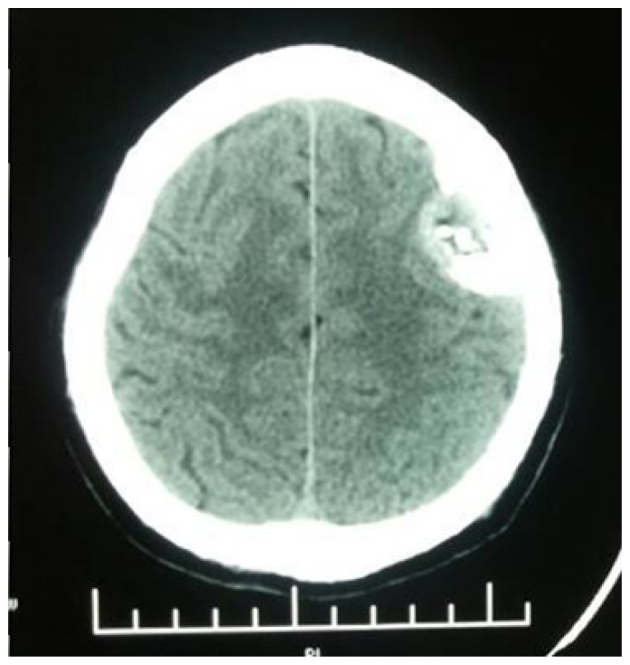
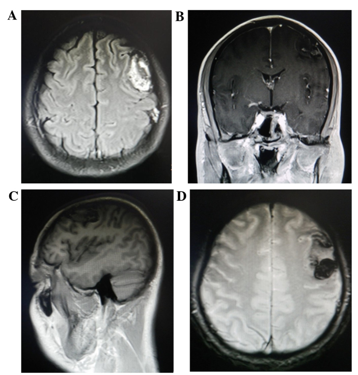
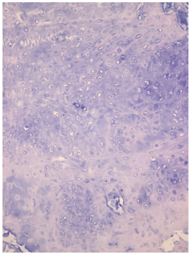
Intracranial chondrosarcoma is a rare malignant cartilage-forming tumor, with only a small number of cases in the posterior cranial fossa reported previously. The present study reports the case of a 40-year-old male patient who was admitted to Tianjin Huanhu Hospital with a progressive headache and dizziness that had lasted for 2 years. Physical and neurological examinations were normal. Radiography of the skull identified an opaque lesion in the left frontal region of the brain. Cranial computed tomography and magnetic resonance imaging revealed a lesion with calcification and homogenous contrast enhancement in the left frontal region. Subsequently, the patient underwent bicoronal craniotomy and gross total resection of the tumor. Pathological examination confirmed the diagnosis of classical intracranial chondrosarcoma. The patient was discharged 10 days after surgery, with no neurological deficit. One month after initial discharge, the patient underwent γ-knife treatment. A follow-up examination 9 months after surgery revealed that the patient was still alive and had returned to work, with no obvious symptoms or evidence of recurrence.
Keywords: central nervous system; chondrosarcoma; intracranial tumor.
Figures
Similar articles
-
Intracranial chondrosarcoma: a case report and review of the literature.Minim Invasive Neurosurg. 2009 Oct;52(5-6):238-41. doi: 10.1055/s-0028-1128117. Epub 2010 Jan 14. Minim Invasive Neurosurg. 2009. PMID: 20077365 Review.
-
A rare tentorial mesenchymal chondrosarcoma in posterior cranial fossa: case report.Neurol Neurochir Pol. 2014;48(4):287-91. doi: 10.1016/j.pjnns.2014.06.001. Epub 2014 Jun 27. Neurol Neurochir Pol. 2014. PMID: 25168329
-
Intracranial extraskeletal mesenchymal chondrosarcoma: case report.Neurosurgery. 2000 Jan;46(1):207-11; discussion 211-2. Neurosurgery. 2000. PMID: 10626952 Review.
-
Primary intracranial extraskeletal myxoid chondrosarcoma: A case report and review of literature.World J Clin Cases. 2022 May 6;10(13):4301-4313. doi: 10.12998/wjcc.v10.i13.4301. World J Clin Cases. 2022. PMID: 35665108 Free PMC article.
-
High-grade intracranial chondrosarcoma presenting with haemorrhage.J Clin Neurosci. 2013 Oct;20(10):1457-60. doi: 10.1016/j.jocn.2012.10.036. Epub 2013 Jun 5. J Clin Neurosci. 2013. PMID: 23746570
Cited by
-
A multicenter retrospective analysis of clinical outcomes of intracranial chondrosarcoma in 26 patients.Sci Rep. 2023 Sep 5;13(1):14647. doi: 10.1038/s41598-023-41378-w. Sci Rep. 2023. PMID: 37669996 Free PMC article.
-
Osteoblastoma in the occipital bone: A case report of a rare tumor in the calvarium.Radiol Case Rep. 2020 Mar 20;15(5):610-614. doi: 10.1016/j.radcr.2020.02.029. eCollection 2020 May. Radiol Case Rep. 2020. PMID: 32215163 Free PMC article.
-
An Unusual Presentation of a Primary Chondrosarcoma of the Cranial Vault.Cureus. 2024 May 16;16(5):e60398. doi: 10.7759/cureus.60398. eCollection 2024 May. Cureus. 2024. PMID: 38883079 Free PMC article.
-
Extra-skeletal intracranial mesenchymal chondrosarcoma: systematic-literature review.Childs Nerv Syst. 2024 Sep;40(9):2723-2733. doi: 10.1007/s00381-024-06452-2. Epub 2024 May 19. Childs Nerv Syst. 2024. PMID: 38762839
-
CT and MRI findings of intracranial extraskeletal mesenchymal chondrosarcoma-a case report and literature review.Transl Cancer Res. 2022 Sep;11(9):3409-3415. doi: 10.21037/tcr-21-2547. Transl Cancer Res. 2022. PMID: 36237268 Free PMC article.
References
LinkOut - more resources
Full Text Sources
Other Literature Sources