. 2016 Nov;12(5):4224-4230.
doi: 10.3892/ol.2016.5153. Epub 2016 Sep 20.

Promotion of metastasis of thyroid cancer cells via NRP-2-mediated induction

Affiliations

Promotion of metastasis of thyroid cancer cells via NRP-2-mediated induction

Dom-Gene Tu et al. Oncol Lett. 2016 Nov.

Abstract

Tumor-node-metastasis is one of the leading causes of morbidity and mortality in thyroid cancer patients. Upregulation of vascular endothelial growth factor-C (VEGF-C) increases the migratory ability of thyroid cancer cells to lymph nodes. Expression of neuropilin-2 (NRP-2), the co-receptor of VEGF-C, has been reported to be correlated with lymph node metastasis in human thyroid cancer. The present study investigated the role of VEGF-C/NRP-2 signaling in the regulation of metastasis of two different types of human thyroid cancer cells. The results indicated that the VEGF-C/NRP-2 axis significantly promoted the metastatic activities of papillary thyroid carcinoma cells through the activation of the mitogen-activated protein kinase (MAPK) kinase (MEK)/extracellular signal-regulated kinase and p38 MAPK signaling cascades. However, neither MEK or p38 MAPK inhibitors produced significant inhibition of the migratory activity and invasiveness regulated by the VEGF-C/NRP-2 axis in follicular thyroid carcinoma cells. Finally, VEGF-C/NRP-2-mediated invasion and migration of thyroid cancer cells required the expression of NRP-2. The present results demonstrate that the promotion of metastasis by VEGF-C is mainly due to the upregulation of NRP-2 in thyroid cancer cells, and this metastatic activity regulated by the VEGF-C/NRP-2 axis provides further insight into the process of tumor metastasis.

Keywords: NRP-2; VEGF-C; lymphangiogenesis; thyroid cancer; tumor metastasis.


Figures

Figure 1.
Figure 1.
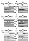
Sequential phosphorylation of ERK and p38 MAPK in response to VEGF-C stimulation in NRP-2-overexpressing thyroid cancer cells. NRP-2-overexpressing (A-C) K1 and (D-F) WRO cells were treated with 100 ng/ml VEGF-C at different time intervals, as indicated. (A) The levels of p-ERK and p-p38 MAPK in K1 cells were measured using western blot analysis. An anti-Myc tag antibody was used to detect the expression of NRP–2. Representative western blots from three independent experiments are shown. (B and C) The results of the densitometric analysis from three independent western blot analyses are shown. (B) p-ERK/total-ERK and (C) p-p38 MAPK/total-p38 MAPK ratios in K1 cells were expressed as the relative variation vs. the basal levels of each single experiment. (D) The levels of p-ERK and p-p38 MAPK in WRO cells were measured using western blot analysis. An anti-Myc tag antibody was used to detect the expression of NRP-2. Representative western blots from three independent experiments are shown. (E and F) The results of the densitometric analysis from three independent western blot analyses are shown. (E) p-ERK/total-ERK and (F) p-p38 MAPK/total-p38 MAPK ratios in WRO cells were expressed as the relative variation vs. the basal levels of each single experiment.***P<0.001. VEGF, vascular endothelial growth factor; NRP-2, neuropilin-2; MAPK, mitogen-activated protein kinase; ERK, extracellular signal-regulated kinase; p-, phosphorylated.
Figure 2.
Figure 2.
Effect of PD, SB and SB2 on the VEGF-C/NRP-2 axis. NRP-2-overexpressing K1 and WRO cells were either pretreated with (A and B) PD (25 µM), (C and D) SB (10 µM), (E and F) SB2 (20 µM) or DMSO for 30 min, and then stimulated with VEGF-C (100 ng/ml) for 10 min. The levels of p-ERK and p-p38 MAPK were measured using western blot analysis. An anti-Myc tag antibody was used to detect the expression of NRP-2. Representative western blots from three independent experiments are shown. VEGF, vascular endothelial growth factor; NRP-2, neuropilin-2; MAPK, mitogen-activated protein kinase; ERK, extracellular signal-regulated kinase; p-, phosphorylated; PD, PD98059; SB, SB203580; SB2, SB202190; DMSO, dimethyl sulfoxide.
Figure 3.
Figure 3.
Effect of the inhibition of the VEGF-C/NRP-2 axis on the metastasis of thyroid cancer cells. NRP-2-overexpressing (A and B) K1 and (C and D) WRO cells were treated with VEGF-C (100 ng/ml) plus or minus PD98059, SB203580 or SB202190. Bar graphs represent the results from (A and C) scratch wound-healing assay (as the mean ± SD of the relative percentages of the wounds gap width) and (B and D) invasion assay (as the mean ± SD of the relative percentages of invading cells) for each treatment from three independent experiments. There was no significant difference in the suppression of VEGF-C/NRP-2-mediated metastasis in NRP-2-overexpressing WRO cells treated with inhibitors plus VEGF-C. *P<0.05; **P<0.01; ***P<0.001. VEGF, vascular endothelial growth factor; NRP-2, neuropilin-2; PD, PD98059; SB, SB203580; SB2, SB202190; DMSO, dimethyl sulfoxide; SD, standard deviation.
Figure 4.
Figure 4.
Effect of the VEGF-C/NRP-2 axis on cell proliferation of thyroid cancer cells. NRP-2-overexpressing (A and B) K1 and (C and D) WRO cells were treated with VEGF-C (100 ng/ml) plus or minus PD98059, SB203580 or SB202190 for (A and C) 8 h or (B and D) 24 h. Bar graphs represent the mean ± standard deviation of the relative percentages of cell proliferation for each treatment from three independent experiments. No significant differences were observed between the different treatments. VEGF, vascular endothelial growth factor; NRP-2, neuropilin-2; MAPK, mitogen-activated protein kinase; PD, PD98059; SB, SB203580; SB2, SB202190; DMSO, dimethyl sulfoxide.
Figure 5.
Figure 5.
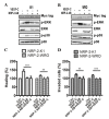
NRP-2 blocking signals suppress VEGF-C-induced metastasis. NRP-2-overexpressing (A) K1 and (B) WRO cells were pretreated with an NRP-2 function-blocking antibody (0.5 µg/ml) for 1.5 h, and then stimulated with VEGF-C (100 ng/ml) for 10 min. The levels of extracellular signal-regulated kinase and p38 mitogen-activated protein kinase phosphorylation were measured using western blot analysis. An anti-Myc tag antibody was used to detect the expression of NRP-2. Representative western blots from three independent experiments are shown. (C) Scratch wound-healing assay and (D) invasion assay were performed to evaluate the effect of NRP-2 blocking signals on cell migration and invasion. Bar graphs represent the mean ± standard deviation of the relative percentages for each treatment from three independent experiments. **P<0.01; ***P<0.001. VEGF, vascular endothelial growth factor; NRP-2, neuropilin-2; MAPK, mitogen-activated protein kinase; ERK, extracellular signal-regulated kinase; p-, phosphorylated; PD, PD98059; SB, SB203580; SB2, SB202190; Ctrl, control; AB, antibody.

References

    1. Fidler IJ. The pathogenesis of cancer metastasis: The ‘seed and soil’ hypothesis revisited. Nat Rev Cancer. 2003;3:453–458. doi: 10.1038/nrc1098. - DOI - PubMed
    1. Joukov V, Pajusola K, Kaipainen A, Chilov D, Lahtinen I, Kukk E, Saksela O, Kalkkinen N, Alitalo K. A novel vascular endothelial growth factor, VEGF-C, is a ligand for the Flt4 (VEGFR-3) and KDR (VEGFR-2) receptor tyrosine kinases. EMBO J. 1996;15:290–298. - PMC - PubMed
    1. Jeltsch M, Kaipainen A, Joukov V, Meng X, Lakso M, Rauvala H, Swartz M, Fukumura D, Jain RK, Alitalo K. Hyperplasia of lymphatic vessels in VEGF-C transgenic mice. Science. 1997;276:1423–1425. doi: 10.1126/science.276.5317.1423. - DOI - PubMed
    1. Amioka T, Kitadai Y, Tanaka S, Haruma K, Yoshihara M, Yasui W, Chayama K. Vascular endothelial growth factor-C expression predicts lymph node metastasis of human gastric carcinomas invading the submucosa. Eur J Cancer. 2002;38:1413–1419. doi: 10.1016/S0959-8049(02)00106-5. - DOI - PubMed
    1. George ML, Tutton MG, Janssen F, Arnaout A, Abulafi AM, Eccles SA, Swift RI. VEGF-A, VEGF-C, and VEGF-D in colorectal cancer progression. Neoplasia. 2001;3:420–427. doi: 10.1038/sj.neo.7900186. - DOI - PMC - PubMed