

Primary Ewing's Sarcoma of the Spine in a Two-Year-Old Boy
- PMID: 27895949
- PMCID: PMC5118528
- DOI: 10.1155/2016/8027137
Primary Ewing's Sarcoma of the Spine in a Two-Year-Old Boy
Abstract
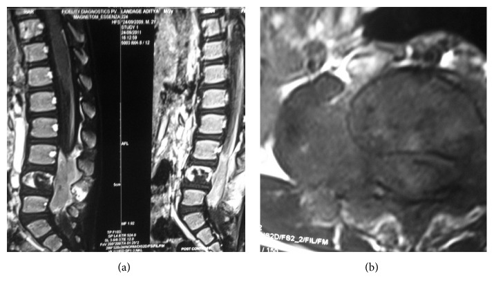
Ewing's Sarcoma (ES) is a highly malignant bone tumour. It may involve any part of the skeleton but the most frequent parts are the ilium and diaphysis of femur and tibia (Alfeeli et al., 2005; Zhu et al., 2012). Primary ES of the spine is extremely rare (Yan et al., 2011). It accounts for only 3.5 to 14.9 percent of all primary bone sarcomas. The age of presentation ranges from 12 to 24 years (median 21 years) (Ferguson, 1999; Sharafuddin et al., 1992; Klimo Jr. et al., 2009). We report an unusual case of primary ES of the spine in a two-year-old boy, who presented to us with paraparesis and features of cauda equina syndrome. MRI scan showed a tumour mass arising from the pedicle of L4 vertebra invading the spinal canal. Tc-99 bone scan showed increased tracer uptake in L4 vertebra and normal tracer uptake elsewhere in the skeleton. After reaching the diagnosis of a space occupying lesion invading the lumber spinal canal, we performed a decompressive laminectomy and a biopsy was sent which confirmed the diagnosis of ES. Immunohistochemistry showed tumour cells staining positive for CD-99 (specific stain for ES). Gene testing showed an EWS-FLI 1 chimera. Surgery was followed by good improvement in motor signs. The child was then referred to a specialized oncotherapy centre for further treatment, radiation, and chemotherapy. To the best of our knowledge, we are the first to report primary ES of the spine at the age of two years.
Conflict of interest statement
The authors declare that there is no conflict of interests.
Figures


References
-
- Ferguson W. S. Chronic leg pain in an adolescent male. Medicine and health, Rhode Island. 1999;82(11):407–409. - PubMed
LinkOut - more resources
Full Text Sources
Other Literature Sources
