. 2016 Oct 3;7(11):4375-4387.
doi: 10.1364/BOE.7.004375. eCollection 2016 Nov 1.

Rapid mesoscale multiphoton microscopy of human skin

Affiliations

Rapid mesoscale multiphoton microscopy of human skin

Mihaela Balu et al. Biomed Opt Express. .

Abstract

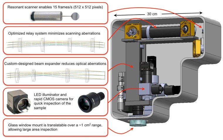
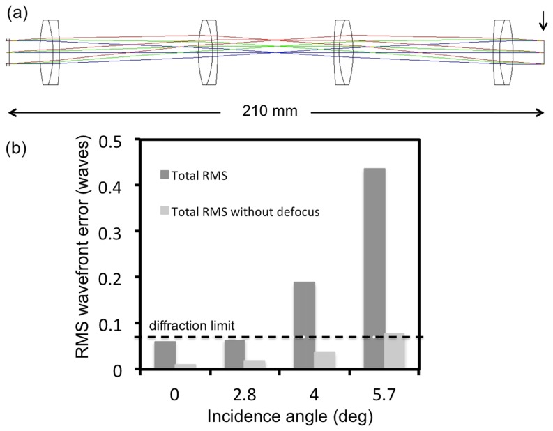
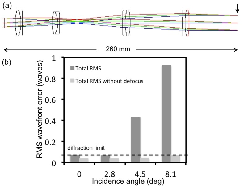
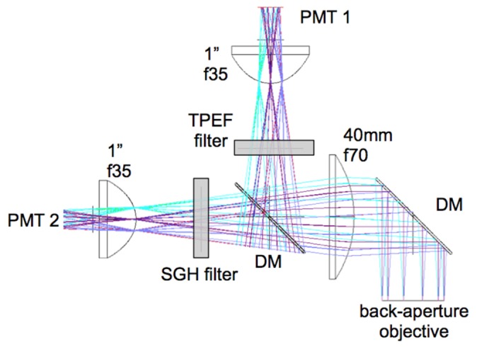
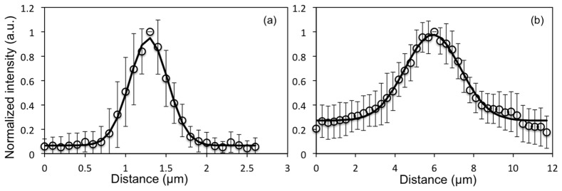
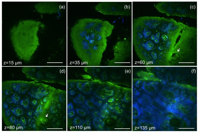
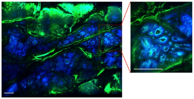
We present a multiphoton microscope designed for mesoscale imaging of human skin. The system is based on two-photon excited fluorescence and second-harmonic generation, and images areas of ~0.8x0.8 mm2 at speeds of 0.8 fps (800x800 pixels; 12 frame averages) for high signal-to-noise ratio, with lateral and axial resolutions of 0.5µm and 3.3µm, respectively. The main novelty of this instrument is the design of the scan head, which includes a fast galvanometric scanner, optimized relay optics, a beam expander and high NA objective lens. Computed aberrations in focus are below the Marechal criterion of 0.07λ rms for diffraction-limited performance. We demonstrate the practical utility of this microscope by ex-vivo imaging of wide areas in normal human skin.

Keywords: (170.2520) Fluorescence microscopy; (180.0180) Microscopy; (180.4315) Nonlinear microscopy.


Figures

Fig. 1
Fig. 1
Schematic and overview of the main elements of the scan head.
Fig. 2
Fig. 2
Relay system-layout and RMS wavefront error. a) optical layout; arrow indicates the image surface at which the aberrations were calculated b) The RMS wavefront error as a function of incidence angle with respect to the diffraction limited value (horizontal line)
Fig. 3
Fig. 3
Beam expander-layout and RMS wavefront error. a) optical layout with the vertical line indicating the surface at which the aberrations are reported b) The RMS wavefront error as a function of incidence angle with respect to the diffraction limited value (horizontal line)
Fig. 4
Fig. 4
Schematic for efficient collection of signals in the epi-direction. Diameter and focal length of the lenses were optimized in ZEMAX for maximum collection of photons. Dichroic mirrors (DM) separate the signal into TPEF and SHG components. Sensitive photomuliplier tubes (PMT) are used.
Fig. 5
Fig. 5
MPM imaging system resolution. Point spread function representing (a) lateral resolution - FWHM = 0.5 µm and (b) axial resolution - FWHM = 3.3 µm. Data were acquired at 800 nm excitation wavelength and represent the fluorescence average of 15 beads. The error bars represent the standard deviation of the fifteen measurements.
Fig. 6
Fig. 6
Ex-vivo human skin imaging-comparison of home-built MPM-based imaging platform with a commercial system using the same objective. (a) Dermo-epidermal junction (DEJ) imaged with the home-built microscope by SHG (blue) and TPEF (green). TPEF signal originates from keratin in the epidermal keratinocytes and from elastin fibers (arrows) in the superficial papillary dermis, while SHG highlights the collagen fibers. (b) A similar location of the DEJ in the skin sample acquired with a commercial Olympus microscope over an area of 370 x 370 µm2 by using the same objective as in the home-built microscope. (c) MPM image of the DEJ acquired with the home-built microscope over an area of 370 x 370 µm2 for comparison with the image in (b) acquired with the Olympus microscope. The image in (c) corresponds to the inset in (a). (d) MPM image of the DEJ corresponding to the inset in (c) showing keratinocytes (full line arrows), elastin fibers (arrow heads) and collagen fibers (dashed line arrows). Images were acquired at 50µm depth in the sample. Scale bar is 100 µm.
Fig. 7
Fig. 7
Ex vivo MPM imaging of human skin at different depths. MPM horizontal sections show images of (a) epidermis (z = 15µm); (b-e) basal cells (green) surrounding dermal papilla (blue) at the dermo-epidermal junction (z = 35µm, 60 µm, 80 µm, 110 µm); arrows indicate hair follicle (c, d); (f) collagen (blue) and elastin (green) fibers in the papillary dermis (z = 135µm). Scale bar is 200 µm.
Fig. 8
Fig. 8
Ex vivo MPM mesoscale imaging of human skin. MPM image of the DEJ in human skin showing keratin in skin folds (green) and fibrilar structure of dermal papilla (collagen fibers-blue; elastin fibers-green). The wide area image (3.1 x 2.5 mm2) includes a mosaic of 20 frames and was acquired in 2 minutes. The image shown as inset was acquired as single frame in 2.4 s. Scale bar is 0.25 mm.

References

    1. Dimitrow E., Ziemer M., Koehler M. J., Norgauer J., König K., Elsner P., Kaatz M., “Sensitivity and specificity of multiphoton laser tomography for in vivo and ex vivo diagnosis of malignant melanoma,” J. Invest. Dermatol. 129(7), 1752–1758 (2009).10.1038/jid.2008.439 - DOI - PubMed
    1. Ulrich M., Klemp M., Darvin M. E., König K., Lademann J., Meinke M. C., “In vivo detection of basal cell carcinoma: comparison of a reflectance confocal microscope and a multiphoton tomograph,” J. Biomed. Opt. 18(6), 061229 (2013).10.1117/1.JBO.18.6.061229 - DOI - PubMed
    1. Balu M., Kelly K. M., Zachary C. B., Harris R. M., Krasieva T. B., König K., Durkin A. J., Tromberg B. J., “Distinguishing between benign and malignant melanocytic nevi by in vivo multiphoton microscopy,” Cancer Res. 74(10), 2688–2697 (2014).10.1158/0008-5472.CAN-13-2582 - DOI - PMC - PubMed
    1. Balu M., Zachary C. B., Harris R. M., Krasieva T. B., König K., Tromberg B. J., Kelly K. M., “In Vivo Multiphoton Microscopy of Basal Cell Carcinoma,” JAMA Dermatol. 151(10), 1068–1074 (2015).10.1001/jamadermatol.2015.0453 - DOI - PMC - PubMed
    1. Balu M., Mazhar A., Hayakawa C. K., Mittal R., Krasieva T. B., König K., Venugopalan V., Tromberg B. J., “In vivo multiphoton NADH fluorescence reveals depth-dependent keratinocyte metabolism in human skin,” Biophys. J. 104(1), 258–267 (2013).10.1016/j.bpj.2012.11.3809 - DOI - PMC - PubMed