Congenital Middle Mesocolic Hernia: A Rare Cause of Neonatal Intestinal Obstruction
- PMID: 27896166
- PMCID: PMC5117281
- DOI: 10.21699/jns.v5i4.371
Congenital Middle Mesocolic Hernia: A Rare Cause of Neonatal Intestinal Obstruction
Abstract
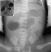
Congenital mesocolic hernia is an extremely rare, but serious cause of intestinal obstruction in children. Given the rarity of this condition, delays in diagnosis and management can have catastrophic consequences. Congenital mesocolic hernias are usually caused by an abnormal rotation of primitive mid-gut and are divided into left and right congenital mesocolic hernias. We report and discuss the clinical and radiological features and management of a neonate with an extremely rare variant, congenital middle mesocolic hernia along with a literature review of this rare condition.
Keywords: Congenital; Middle mesocolic hernia; Neonatal intestinal obstruction.
Figures


References
-
- Ghahremani GG. Internal abdominal hernias. Surg Clin North Am. 1984; 64:393-406. - PubMed
-
- Newsom BD, Kukora JS. Congenital and acquired internal hernias: unusual causes of small bowel occlusion. Am J Surg. 1986;152:279–85. - PubMed
-
- Tong RS, Sengupta S, Tjandra JJ. Left paraduodenal hernia: case report and review of the literature. ANZ J Surg. 2002; 72:69–71. - PubMed
-
- Martin LC, Merkle EM, Thompson WM. Review of internal hernias: radiographic and clinical findings. AJR Am J Roentgenol. 2006; 186:703-17. - PubMed
-
- Page MP, Ricca RL, Resnick AS, Puder M, Fishman SJ. Newborn and toddler intestinal obstruction owing to congenital mesenteric defects. J Pediatr Surg. 2008; 43:755-8. - PubMed
Publication types
LinkOut - more resources
Full Text Sources
Other Literature Sources
