Uterine arteriovenous malformation with positive serum beta-human chorionic gonadotropin: Embolization of both uterine arteries and extra-uterine feeding arteries
- PMID: 27896262
- PMCID: PMC5120079
- DOI: 10.5468/ogs.2016.59.6.554
Uterine arteriovenous malformation with positive serum beta-human chorionic gonadotropin: Embolization of both uterine arteries and extra-uterine feeding arteries
Abstract
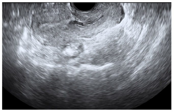
The incidence of uterine arteriovenous malformation (AVM) is rare. However, it is clinically significant in that it can cause life-threatening vaginal bleeding. We report a case of a large uterine AVM with positive serum beta-human chorionic gonadotropin. A presumptive diagnosis was made; a uterine AVM accompanied by, early pregnancy or retained product of conception. Because this uterine AVM was extensive, transcatheter arterial embolization of both uterine arteries and extra-uterine feeding arteries was performed. Three months after undergoing transcatheter arterial embolization, complete resolution of the uterine AVM was confirmed without major complication.
Keywords: Arteriovenous malformations; Beta-human chorionic gonadotropin; Transcatheter arterial embolization.
Conflict of interest statement
No potential conflict of interest relevant to this article was reported.
Figures

Similar articles
-
Uterine Arteriovenous Malformation With Raised Serum Beta-Human Chorionic Gonadotropin (β-hCG) Levels: A Diagnostic and Therapeutic Dilemma.Cureus. 2024 Jul 10;16(7):e64209. doi: 10.7759/cureus.64209. eCollection 2024 Jul. Cureus. 2024. PMID: 39130918 Free PMC article.
-
Successful treatment of uterine arteriovenous malformation with percutaneous embolization.Taiwan J Obstet Gynecol. 2007 Mar;46(1):60-3. doi: 10.1016/S1028-4559(08)60109-6. Taiwan J Obstet Gynecol. 2007. PMID: 17389192
-
Effect of gonadotropin-releasing hormone agonist on a uterine arteriovenous malformation.Obstet Gynecol. 2006 Sep;108(3 Pt 2):751-3. doi: 10.1097/01.AOG.0000191584.28717.2c. Obstet Gynecol. 2006. PMID: 17018490
-
Use of three-dimensional power Doppler sonography in the diagnosis of uterine arteriovenous malformation and follow-up after uterine artery embolization: Case report and brief review of literature.J Clin Ultrasound. 2015 Jun;43(5):327-34. doi: 10.1002/jcu.22210. Epub 2014 Jul 18. J Clin Ultrasound. 2015. PMID: 25042165 Review.
-
Transcatheter embolization of uterine arteriovenous malformation: report of 2 cases and review of literature.J Minim Invasive Gynecol. 2011 Nov-Dec;18(6):812-9. doi: 10.1016/j.jmig.2011.07.007. J Minim Invasive Gynecol. 2011. PMID: 22024270 Review.
Cited by
-
Uterine arteriovenous malformation with repeated vaginal bleeding after dilatation and curettage.Obstet Gynecol Sci. 2019 Mar;62(2):142-145. doi: 10.5468/ogs.2019.62.2.142. Epub 2019 Mar 4. Obstet Gynecol Sci. 2019. PMID: 30918884 Free PMC article.
-
Retained Placenta Percreta with Acquired Uterine Arteriovenous Malformation-Case Report and Short Review of the Literature.Diagnostics (Basel). 2022 Apr 5;12(4):904. doi: 10.3390/diagnostics12040904. Diagnostics (Basel). 2022. PMID: 35453952 Free PMC article.
-
Uterine Arteriovenous Fistula with Concomitant Pelvic Varicocele: Endovascular Embolization with Onyx-18®.Case Rep Vasc Med. 2017;2017:3548271. doi: 10.1155/2017/3548271. Epub 2017 Nov 22. Case Rep Vasc Med. 2017. PMID: 29359062 Free PMC article.
References
-
- Timmerman D, Van den Bosch T, Peeraer K, Debrouwere E, Van Schoubroeck D, Stockx L, et al. Vascular malformations in the uterus: ultrasonographic diagnosis and conservative management. Eur J Obstet Gynecol Reprod Biol. 2000;92:171–178. - PubMed
-
- Kwon JH, Kim GS. Obstetric iatrogenic arterial injuries of the uterus: diagnosis with US and treatment with transcatheter arterial embolization. Radiographics. 2002;22:35–46. - PubMed
-
- O'Brien P, Neyastani A, Buckley AR, Chang SD, Legiehn GM. Uterine arteriovenous malformations: from diagnosis to treatment. J Ultrasound Med. 2006;25:1387–1392. - PubMed
-
- Grivell RM, Reid KM, Mellor A. Uterine arteriovenous malformations: a review of the current literature. Obstet Gynecol Surv. 2005;60:761–767. - PubMed
-
- Kelly SM, Belli AM, Campbell S. Arteriovenous malformation of the uterus associated with secondary postpartum hemorrhage. Ultrasound Obstet Gynecol. 2003;21:602–605. - PubMed
Publication types
LinkOut - more resources
Full Text Sources
Other Literature Sources