Review
. 2016 Nov 15:4:87.
doi: 10.3389/fbioe.2016.00087. eCollection 2016.

In Vitro Bone Cell Models: Impact of Fluid Shear Stress on Bone Formation

Affiliations
Review

In Vitro Bone Cell Models: Impact of Fluid Shear Stress on Bone Formation

Claudia Wittkowske et al. Front Bioeng Biotechnol. .

Abstract

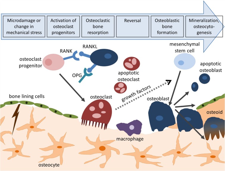
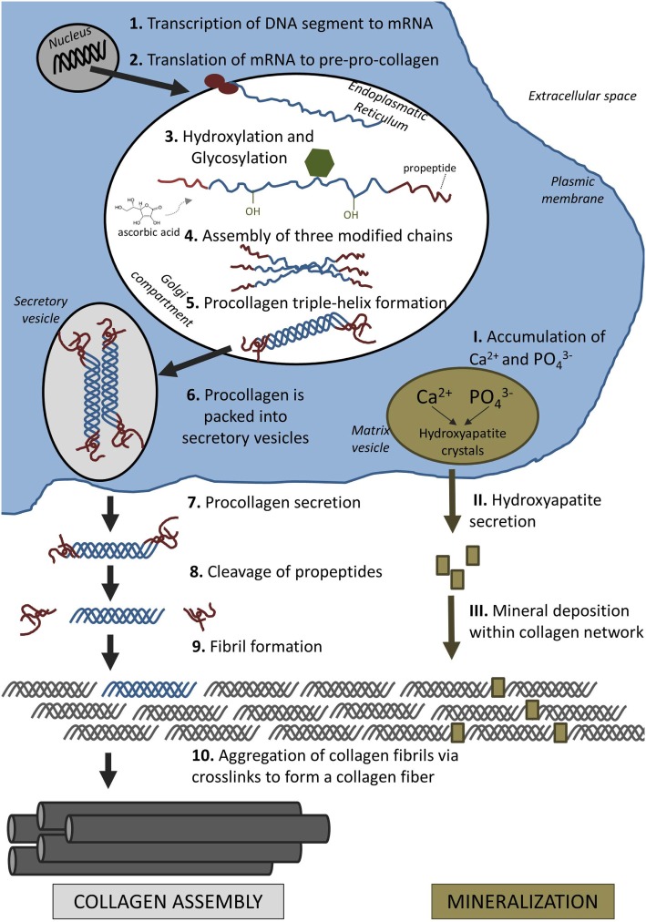
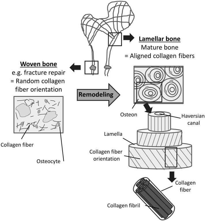
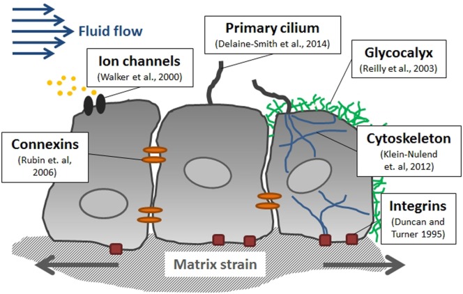
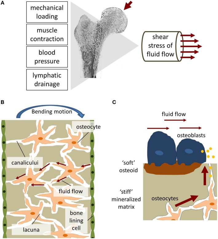
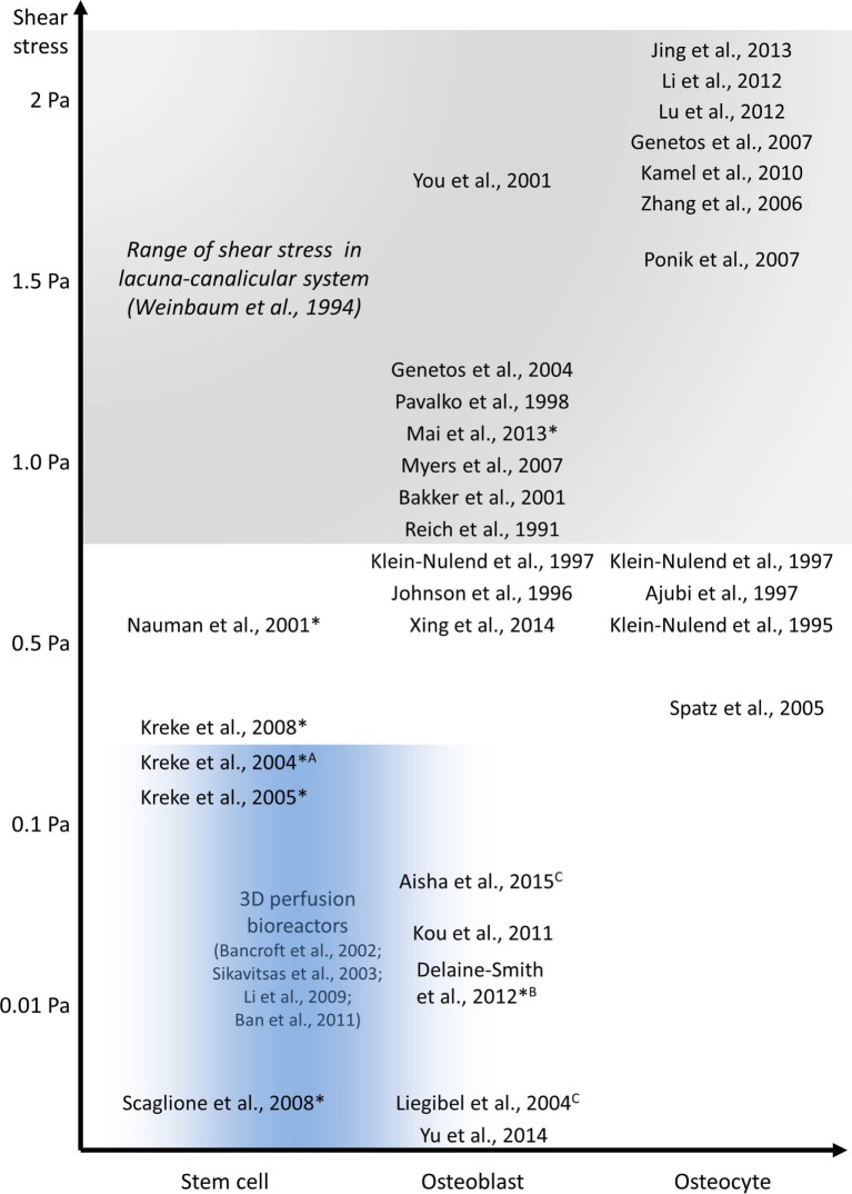
This review describes the role of bone cells and their surrounding matrix in maintaining bone strength through the process of bone remodeling. Subsequently, this work focusses on how bone formation is guided by mechanical forces and fluid shear stress in particular. It has been demonstrated that mechanical stimulation is an important regulator of bone metabolism. Shear stress generated by interstitial fluid flow in the lacunar-canalicular network influences maintenance and healing of bone tissue. Fluid flow is primarily caused by compressive loading of bone as a result of physical activity. Changes in loading, e.g., due to extended periods of bed rest or microgravity in space are associated with altered bone remodeling and formation in vivo. In vitro, it has been reported that bone cells respond to fluid shear stress by releasing osteogenic signaling factors, such as nitric oxide, and prostaglandins. This work focusses on the application of in vitro models to study the effects of fluid flow on bone cell signaling, collagen deposition, and matrix mineralization. Particular attention is given to in vitro set-ups, which allow long-term cell culture and the application of low fluid shear stress. In addition, this review explores what mechanisms influence the orientation of collagen fibers, which determine the anisotropic properties of bone. A better understanding of these mechanisms could facilitate the design of improved tissue-engineered bone implants or more effective bone disease models.

Keywords: bone remodeling; collagen orientation; fluid shear stress; osteoblast; osteocyte.


Figures

Figure 1
Figure 1
Bone remodeling cycle. Bone remodeling is initiated by microcracks or changes in mechanical loading and consists of four consecutive steps: activation, resorption, reversal, and formation. Activation of osteoclasts is controlled through the RANK/RANKL/OPG pathway. Following bone deposition, osteoblasts can differentiate to osteocytes (osteocytogenesis), turn to bone-lining cells, or enter apoptosis.
Figure 2
Figure 2
Formation of extracellular matrix: collagen assembly and mineralization.
Figure 3
Figure 3
Woven and lamellar bone. Two types of bone can be distinguished based on the orientation of collagen fibers within the bone matrix. Woven bone (left) consists of randomly oriented collagen fibrils and exists during fetal development and fracture repair. Following bone remodeling, most mature bone can be characterized as lamellar bone (right). Lamellar bone is characterized by highly orientated collagen fibers arranged in lamellae and osteons. The alternating orientation of the collagen lamellae results in significantly higher strength of lamellar bone compared to woven bone.
Figure 4
Figure 4
Mediators of mechanotransduction in bone. Several cellular structures have been identified in bone cells which contribute to sensing mechanical stimuli including fluid flow-induced shear stress and matrix strain.
Figure 5
Figure 5
Fluid shear stress in bone. (A) Fluid flow in bone can be generated through mechanical loading, muscle contraction, blood pressure, and lymphatic drainage. (B) Bending of bone generates tension and compression forces which initiates interstitial fluid flow [adapted from Duncan and Turner (1995)]. (C) The biomechanical environment experienced by osteoblasts and osteocytes differs regarding flow velocities and matrix stiffness.
Figure 6
Figure 6
In vitro studies investigating the effect of fluid shear stress on osteoblast and osteocyte behavior. All experiments were performed using PPFC, exceptions: Aradial flow chamber, Brocking platform, Corbital shaker, and 3D perfusion bioreactor experiments in blue area. Long-term experiments are indicated with *.

References

    1. Aisha M. D., Nor-Ashikin M. N. K., Sharaniza A. B. R., Nawawi H., Froemming G. R. A. (2015). Orbital fluid shear stress promotes osteoblast metabolism, proliferation and alkaline phosphates activity in vitro. Exp. Cell Res. 337, 87–93.10.1016/j.yexcr.2015.07.002 - DOI - PubMed
    1. Ajubi N. E., Klein-Nulend J., Alblas M. J., Burger E. H., Nijweide P. J. (1999). Signal transduction pathways involved in fluid flow-induced PGE2 production by cultured osteocytes. Am. J. Physiol. Endocrinol. Metab. 276, E171–E178. - PubMed
    1. Al Nazer R., Lanovaz J., Kawalilak C., Johnston J. D., Kontulainen S. (2012). Direct in vivo strain measurements in human bone-a systematic literature review. J. Biomech. 45, 27–40.10.1016/j.jbiomech.2011.08.004 - DOI - PubMed
    1. Anderson E. J., Falls T. D., Sorkin A. M., Knothe Tate M. L. (2006). The imperative for controlled mechanical stresses in unraveling cellular mechanisms of mechanotransduction. Biomed. Eng. Online 5, 27.10.1186/1475-925X-5-27 - DOI - PMC - PubMed
    1. Anderson H. C. (1995). Molecular biology of matrix vesicles. Clin. Orthop. Relat. Res. 314, 266–280. - PubMed