. 2016 Dec;27(6):209-218.
doi: 10.1089/hgtb.2016.120.

A Rapid Cell Expansion Process for Production of Engineered Autologous CAR-T Cell Therapies

Affiliations

A Rapid Cell Expansion Process for Production of Engineered Autologous CAR-T Cell Therapies

Tangying Lily Lu et al. Hum Gene Ther Methods. 2016 Dec.

Erratum in

  • Correction to: Hum Gene Ther Methods 2016;27:209-218.
    [No authors listed] [No authors listed] Hum Gene Ther Methods. 2017 Apr;28(2):100. doi: 10.1089/hgtb.2016.120.correx. Epub 2017 Feb 22. Hum Gene Ther Methods. 2017. PMID: 28394690 Free PMC article. No abstract available.

Abstract

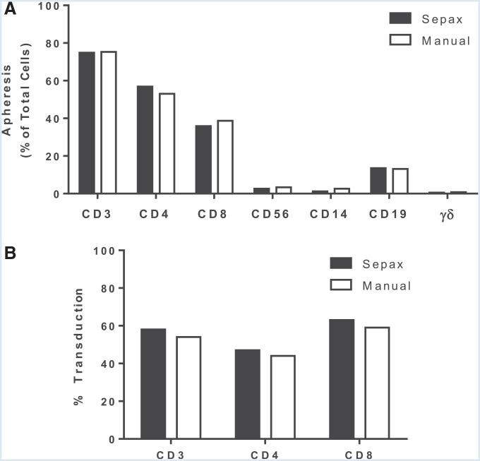
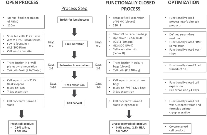
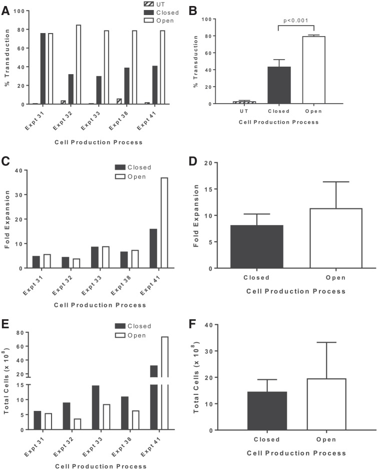
The treatment of B-cell malignancies by adoptive cell transfer (ACT) of anti-CD19 chimeric antigen receptor T cells (CD19 CAR-T) has proven to be a highly successful therapeutic modality in several clinical trials.1-6 The anti-CD19 CAR-T cell production method used to support initial trials relied on numerous manual, open process steps, human serum, and 10 days of cell culture to achieve a clinical dose.7 This approach limited the ability to support large multicenter clinical trials, as well as scale up for commercial cell production. Therefore, studies were completed to streamline and optimize the original National Cancer Institute production process by removing human serum from the process in order to minimize the risk of viral contamination, moving process steps from an open system to functionally closed system operations in order to minimize the risk of microbial contamination, and standardizing additional process steps in order to maximize process consistency. This study reports a procedure for generating CD19 CAR-T cells in 6 days, using a functionally closed manufacturing process and defined, serum-free medium. This method is able to produce CD19 CAR-T cells that are phenotypically and functionally indistinguishable from cells produced for clinical trials by the previously described production process.

Keywords: GMP; anti-CD19 CAR; closed system; cryopreservation; expansion; transduction.


Conflict of interest statement

M.B. has employment and equity ownership in Kite Pharma. O.P. is a former KITE Pharma employee with no outstanding financial interests. No competing financial interests exist for the remaining authors.

Figures

<b>Figure 1.</b>
Figure 1.
Comparison of peripheral blood mononuclear cells (PBMCs) isolated from apheresis using Sepax II or manual Ficoll separation. (A) Apheresis from a healthy donor was split equally and processed using the Sepax II density gradient-based preparation with the kit CS-900 from Biosafe or by manual Ficoll separation. The following lymphocyte cell subpopulations were analyzed by fluorescence-activated cell sorting (FACS): total lymphocytes (CD3+), natural killer (NK; CD3CD56+), CD19+ cells, gd T cells, and, within the CD3+ T cells, the percentages of T helper (CD3+CD4+) and CTL (CD3+CD8+). (B) T cells isolated from both procedures were activated with soluble OKT3 and transduced with PG13-CD19-H3 vector. The vector transduction efficiency was measured by FACS. Data are presented from a single experiment and are representative of at least three independent experiments.
<b>Figure 2.</b>
Figure 2.
Optimization of T-cell growth in serum-free medium. Lymphocytes were activated with OKT3 for 2 days and expanded in media containing interleukin-2 (IL-2) for 6 days. During the expansion phase, cells were split to 0.5e6/mL when reaching a density of 2 × 106 cells/mL. (A) The number of total viable cells cultured in the various medium in the absence of serum, or (B) in the same medium supplemented with 5% T-cell serum replacement (TCSR). Data are from three independent experiments represented as the mean ± the standard error of the mean (SEM).
<b>Figure 3.</b>
Figure 3.
Comparison of CD19 chimeric antigen receptor (CAR) T cell transduction by spinoculation or in static bags. Apheresis from three subjects was processed either manually or using the Sepax II, and PBMCs were activated with OKT3 in AIM V medium containing 5% human serum and 300 IU of IL-2 for 2 days. Cells were then transduced with PG13-CD19-H3 Vector by spinoculation in six-well plates (open) or in static culture using Origen PermaLife PL07 bags (closed). (A) Four days post transduction, cells were analyzed by FACS for the percentages of CD3+ and CD4+ or CD8+ (of the CD3+) transduced cells. (B) CD19 CAR-T cells were co-cultured with CD19+ target cells (Toledo and Nalm6) or CD19 (K562-NGFR and CEM). The next day, supernatants were collected, and interferon gamma (IFN-γ) was detected by enzyme-linked immunosorbent assay. The data represent three independent experiments and are presented as mean ± SEM.
<b>Figure 4.</b>
Figure 4.
Optimization of closed transduction process for the manufacture of CD19 CAR-T cells. Lymphocytes were isolated from subject apheresis products using the Sepax II and stimulated as previously described with the following changes. (A) After stimulation, PBMCs were seeded at the indicated cell density and transduced in bags as previously described. The transduction efficiency was measured by FACS staining for CD19 CAR expression 4 days post transduction. (B) Origen cell culture bags were coated with retronectin at the indicated concentrations. The next day, retronectin was removed and the bags were blocked with 2.5% human serum albumin (HSA) before vector loading. OKT3 activated-PBMCs from three patients (1 × 106 cells/mL) were added to separate bags, and transduction efficiency was measured by FACS. (C) To evaluate the need for a blocking step following retronectin coating of the bag, bags were coated with 10 μg/mL of retronectin and blocked with either 2.5% HSA or HBSS and then loaded with retroviral vector. No significant difference in transduction was observed by the addition of a blocking step. However, removal of the retroviral vector prior to the T cell transduction did significantly reduce the level of CD19 CAR expression (p = 0.004, stippled bar). (D) In the closed transduction process (control), the bag is coated with retronectin overnight followed by a wash-and-block step prior to vector loading. In an attempt to simplify the process, the block step prior to transduction was removed and the addition of retronectin and vector prior to the addition of cells was combined. In a separate effort, the addition of retronectin, vector, and cells was combined into a single step. All other conditions were the same. All data are representative of at least three independent experiments. Data are presented as mean ± SEM.
<b>Figure 5.</b>
Figure 5.
Manufacturing scheme for the functionally closed production of CD19 CAR-T cells. An overview of the 6-day cell production process developed at the Surgery Branch of the National Cancer Institute. The schematic shows a comparison between the 10-day open spinoculation process for the administration of a non-cryopreserved cell product and the new 6-day closed-cell production process followed by cryopreservation of the final cell product. Optimizations achieved at each process step are indicated.
<b>Figure 6.</b>
Figure 6.
Comparison of an open versus functionally closed production system for the manufacture of CD19 CAR-T cells. To evaluate the efficacy of the production process with closed-unit operations, five engineering runs were performed at scale and compared directly to cells manufactured using the open spinoculation process. (A and B) The transduction of PBMCs with anti-CD19 CAR viral vectors in a closed bag system (closed) was compared to the previous Surgery Branch (open) plate transduction platform. In both cases, 1 × 109 PBMCs were stimulated with soluble OKT3 for 2 days, followed by transduction of PBMCs at a density of 0.5 × 106 cells/mL with a 2× diluted vector supernatant. Transduction was significantly higher for cell manufactured using the open system (p < 0.001). (C and D) The comparison of the cell expansion at the end of the 6 days. (E and F) The comparison of total cell numbers between open and closed production process over 6 days. The data are representative of five engineering runs at scale. Individual experiments (A, C, and E) are plotted, as well as summary data (B, D, and F) for each parameter evaluated. Summary data are presented as the mean ± SEM.

References

    1. Howlader N, Noone AM, Krapcho M, et al. . SEER Cancer Statistics Review, 1975–2013. Bethedsda, MD: National Cancer Institute, 2016
    1. Gisselbrecht C, Glass B, Mounier N, et al. . Salvage regimens with autologous transplantation for relapsed large B-cell lymphoma in the rituximab era. J Clin Oncol 2010;28:4184–4190 - PMC - PubMed
    1. Kenkre VP, Smith SM. Management of relapsed diffuse large B-cell lymphoma. Curr Oncol Rep 2008;10:393–403 - PubMed
    1. Sehn LH, Fenske TS, Laport GG. Follicular lymphoma: prognostic factors, conventional therapies, and hematopoietic cell transplantation. Biol Blood Marrow Transplant 2012;18:S82–91 - PubMed
    1. Tedder TF, Isaacs CM. Isolation of cDNAs encoding the CD19 antigen of human and mouse B lymphocytes. A new member of the immunoglobulin superfamily. J Immunol 1989;143:712–717 - PubMed

Publication types