. 2016 Nov 29:7:13668.
doi: 10.1038/ncomms13668.

Truncation and constitutive activation of the androgen receptor by diverse genomic rearrangements in prostate cancer

Affiliations

Truncation and constitutive activation of the androgen receptor by diverse genomic rearrangements in prostate cancer

Christine Henzler et al. Nat Commun. .

Abstract

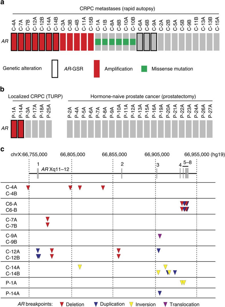
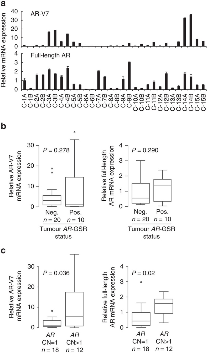
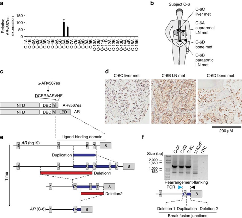
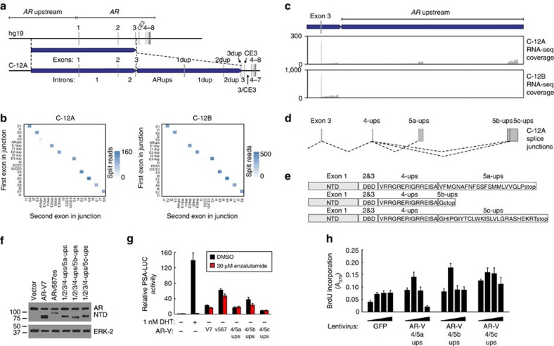
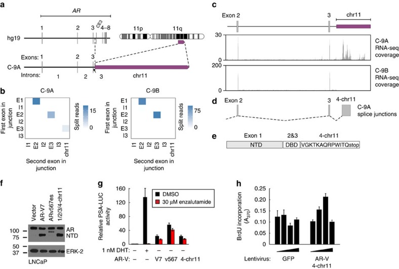
Molecularly targeted therapies for advanced prostate cancer include castration modalities that suppress ligand-dependent transcriptional activity of the androgen receptor (AR). However, persistent AR signalling undermines therapeutic efficacy and promotes progression to lethal castration-resistant prostate cancer (CRPC), even when patients are treated with potent second-generation AR-targeted therapies abiraterone and enzalutamide. Here we define diverse AR genomic structural rearrangements (AR-GSRs) as a class of molecular alterations occurring in one third of CRPC-stage tumours. AR-GSRs occur in the context of copy-neutral and amplified AR and display heterogeneity in breakpoint location, rearrangement class and sub-clonal enrichment in tumours within and between patients. Despite this heterogeneity, one common outcome in tumours with high sub-clonal enrichment of AR-GSRs is outlier expression of diverse AR variant species lacking the ligand-binding domain and possessing ligand-independent transcriptional activity. Collectively, these findings reveal AR-GSRs as important drivers of persistent AR signalling in CRPC.


Conflict of interest statement

S.R.P. and S.M.D. have served as a paid consultant/advisor for Medivation/Astellas. The remaining authors declare no competing financial interests.

Figures

Figure 1
Figure 1. Frequent and diverse AR gene structural rearrangements in CRPC metastases.
(a) Oncoprint illustrating the presence of AR amplification, missense mutations or AR-GSRs in 30 metastases from 15 rapid autopsy subjects. (b) Oncoprint illustrating the presence of AR amplification or AR-GSRs in 6 localized CRPC specimens obtained by TURP and 21 hormone-naive prostate cancer specimens. (c) Map of AR-GSR breakpoint locations within the AR gene body for nine AR-GSR patients. Coloured triangles represent break fusion junction locations, with the colour specifying whether that break fusion junction was due to a discrete deletion (red), duplication (blue), inversion (yellow) or translocation (purple) event. Genome coordinates are genome build GRCh37/hg19. Locations of AR exons 1–8 are shown.
Figure 2
Figure 2. Association of AR-V7 mRNA expression levels with AR gene copy number.
(a) mRNA expression of AR-V7 (top) and full-length AR (bottom) was assessed by quantitative RT–PCR. Levels are shown relative to a housekeeping gene (RPL13A), with expression in tumour C-1A set to 1. Data represent mean±s.d. from three repeated experiments each consisting of independent RT and PCR reactions from the same original RNA sample (n=3). Relative mRNA expression levels of AR-V7 (left) or full-length AR (right) were compared in tumours that (b) were positive (Pos.) or negative (Neg.) for an AR gene structural rearrangement (AR-GSR) event or (c) had AR gene copy number (CN) ≥1. Centre lines show the medians; box limits indicate the 25th and 75th percentiles; whiskers extend 1.5 times the interquartile range from the 25th and 75th percentiles and outliers are represented by dots. P-values were determined by one-tailed Mann–Whitney U-test.
Figure 3
Figure 3. Outlier expression of ARv567es in subject C-6 harbouring a tumour clone with a complex AR-GSR.
(a) mRNA expression of ARv567es was assessed by quantitative RT–PCR. Levels are shown relative to a housekeeping gene (RPL13A), with expression in tumour C-1A set to 1. Data represent mean±s.d. from three repeated experiments, each consisting of independent RT and PCR reactions from the same original RNA sample (n=3). (b) Location of metastatic sites analysed for subject C-6. (c) Location and sequence of the unique epitope recognized by a rabbit monoclonal antibody specific for ARv567es. (d) Three tumour sites from subject C-6 were stained by immunohistochemistry with an antibody specific for ARv567es, revealing strong nuclear expression. (e) Model for occurrence of a multi-step rearrangement affecting the sole AR gene copy in subject C-6. (f) PCR validating existence of the multi-step AR rearrangement illustrated in e in genomic DNA from three tumour sites from subject C-6. Sanger sequencing of PCR products revealed that all three break fusion junction signatures (duplication and deletions 1 and 2) existed on the same DNA molecule.
Figure 4
Figure 4. An AR-GSR in tumour C-12A drives expression of a set of duplication-dependent AR-V mRNAs.
(a) Schematic of AR exon and intron organization resulting from the 379 kb tandem duplication in tumour C-12A. (b) Heatmaps representing numbers of RNA-seq reads from tumours C-12A and C-12B spanning exon/exon, exon/intron and intron/intron boundaries of the AR architecture defined in a. Each exon and intron was considered as a discrete ‘bin', with each pixel representing counts of RNA-seq splice junctions starting in the first bin (first exon in junction) and ending in the second bin (second exon in junction). (c) RNA-seq read coverage from tumour C-12A within the AR upstream region that becomes situated downstream of AR exon 3 as a result of the 379 kb tandem duplication. RNA-seq read coverage for tumour C-12B, which is negative for this tandem duplication is shown as a control. (d) Novel AR splice junctions occurring in tumour C-12A. Exons are named ‘4-ups', ‘5a-ups', ‘5b-ups' and ‘5c-ups' to denote their splicing order (4 or 5) in the AR-V mRNA and their genomic locations upstream from AR in the reference genome architecture. (e) Translation of three novel AR-V mRNA species uniquely expressed in tumour C-12A consisting of the AR NTD, DBD and unique COOH-termini. (f,g) LNCaP cells were transfected with a PSA promoter/enhancer-luciferase reporter and expression vectors encoding AR-V7, ARv567es or AR-Vs from tumour C-12A. Cells were treated with 1 nM dihydrotestosterone (DHT), 30 μM enzalutamide (enz) or DMSO (vehicle control) as indicated and subjected to (f) western blotting with antibodies specific for the AR NTD or ERK-2 (loading control), or (g) Luciferase assay. Data represent mean±s.e.m. from three biological replicate experiments, each performed in duplicate (n=6). (h) LNCaP cells were infected with a range of titres of lentivirus encoding GFP (control) or AR-Vs from tumour C-12A and assayed for proliferation by BrdU incorporation assay. Data represent mean±s.e.m. from two biological replicate experiments, each performed in triplicate (n=6).
Figure 5
Figure 5. An AR-GSR in tumour C-9A drives expression of a translocation-dependent AR-V mRNA.
(a) Schematic of AR exon and intron organization resulting from the AR:chr11 translocation in tumour C-9A. (b) Heatmaps representing numbers of RNA-seq reads from tumours C-9A and C-9B spanning exon/exon, exon/intron and intron/intron boundaries of the AR architecture defined in a. Each exon and intron was considered as a discrete ‘bin', with each pixel representing counts of RNA-seq splice junctions starting in the first bin (first exon in junction) and ending in the second bin (second exon in junction). (c) RNA-seq read coverage from tumour C-9A within the chromosome 11 region that becomes situated downstream of AR exon 3 as a result of the translocation. RNA-seq read coverage from tumour C-9B, which is negative for this rearrangement, is shown as a control. (d) Novel AR splice junctions occurring in tumour C-9A. (e) Translation of a novel AR-V mRNA species uniquely expressed in tumour C-9A consisting of the AR NTD, DBD and a unique COOH terminus. (f,g) LNCaP cells were transfected with a PSA promoter/enhancer-luciferase reporter and expression vectors encoding AR-Vs as indicated. The AR-V encoded by splicing of a novel AR exons from chromosome 11 is indicated in bold. Cells were treated with dihydrotestosterone (DHT), enzalutamide (enz) or DMSO (vehicle control) as indicated and subjected to (f) western blotting with antibodies specific for the AR NTD or ERK-2 (loading control), or (g) Luciferase assay. Data represent mean±s.e.m. from three biological replicate experiments, each performed in technical duplicate (n=6). (h) LNCaP cells were infected with a range of titres of lentivirus encoding GFP (control) or the unique AR-V expressed in tumour C-9A and assayed for proliferation by BrdU incorporation assay. Data represent mean±s.e.m. from two biological replicate experiments, each performed in triplicate (n=6).

References

    1. Grasso C. S. et al.. The mutational landscape of lethal castration-resistant prostate cancer. Nature 487, 239–243 (2012). - PMC - PubMed
    1. Gundem G. et al.. The evolutionary history of lethal metastatic prostate cancer. Nature 520, 353–357 (2015). - PMC - PubMed
    1. Robinson D. et al.. Integrative clinical genomics of advanced prostate cancer. Cell 161, 1215–1228 (2015). - PMC - PubMed
    1. Taylor B. S. et al.. Integrative genomic profiling of human prostate cancer. Cancer Cell 18, 11–22 (2010). - PMC - PubMed
    1. Beltran H. et al.. Divergent clonal evolution of castration-resistant neuroendocrine prostate cancer. Nat. Med. 22, 298–305 (2016). - PMC - PubMed

Publication types