. 2016 Nov 29:6:37867.
doi: 10.1038/srep37867.

Hydroxychavicol: A phytochemical targeting cutaneous fungal infections

Affiliations

Hydroxychavicol: A phytochemical targeting cutaneous fungal infections

Intzar Ali et al. Sci Rep. .

Abstract

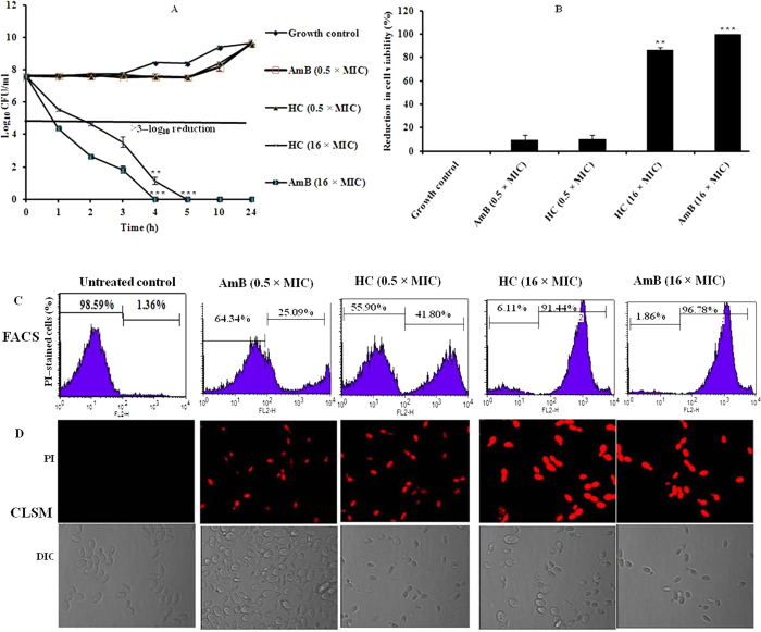
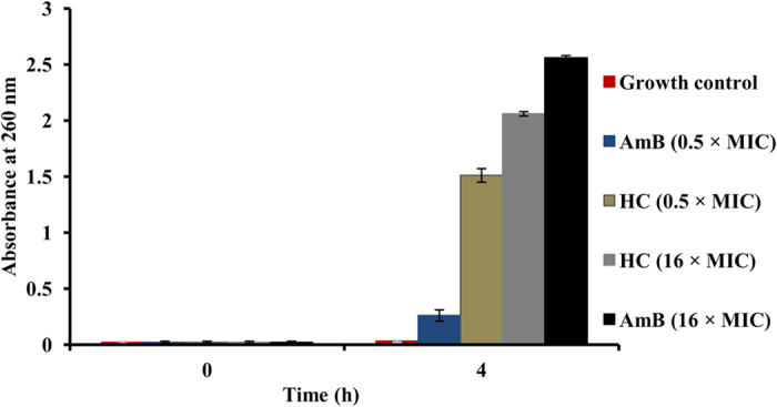
The present study was designed to investigate the potency of hydroxychavicol on selected cutaneous human pathogenic fungi by the use of in vitro and in vivo assays and mechanistic characterization along with toxicological effects. Hydroxychavicol consistently displayed a fungicidal effect against all fungal species tested. Inoculum concentrations over the range of 104 to 107 CFU/ml did not significantly alter its antifungal potential and time-kill curve results revealed concentration-dependent killing. It also inhibited the growth of biofilm generated by Trichophyton mentagrophytes and Candida parapsilosis and reduced the preformed biofilms. Hydroxychavicol was highly effective in the treatment, and mycological eradication of an experimentally induced topical infection model of dermatophytosis (tinea corporis) and cutaneous candidiasis in guinea pigs, respectively. The mode of action of hydroxychavicol appears to originate from the disruption of cell membrane integrity. Administration of hydroxychavicol in mice at 500 mg per kg of body weight by orally produced no overt toxicity. The retention capacity of hydroxychavicol in vitro, in the presence of keratin has attributed to its in vivo effectiveness in the guinea pig model of topical infections. Furthermore, it is suggestive of its potential use as phytochemical for topical use in cutaneous fungal infections.


Figures

Figure 1
Figure 1
Time–kill curve plots of hydroxychavicol (HC) against Candida parapsilosis ATCC 200954 (A) and the mixture of germinated as well as ungerminated microconidia of Trichophyton mentagrophytes var. interdigitale ATCC 9533 (B) following exposure of 1 to 16 times the MICs. The cultures were incubated in RPMI 1640 medium buffered to a pH of 7.0 with 0.165 M MOPS buffer (CLSI). The lower limit of accurate and reproducible detectable colony counts was 10 CFU/ml. The solid lines represent a >99.9% growth reduction compared with that of initial inoculums added at the start of the experiment (fungicidal effect). Each time point represents as the mean log10 ± standard deviation of two different experiments performed in duplicate with similar results of HC–treated group each time. *P < 0.05; **P < 0.005; ***P < 0.0001 (Student’s t–test) were considered to be statistically significant when compared with the untreated growth control.
Figure 2
Figure 2. Effect of hydroxychavicol (HC) on the cell membrane permeability of Candida parapsilosis ATCC 200954, measured by uptake of propidium iodide (PI).
The mid–log growth phase culture at a cell density of approximately 4 × 107 CFU/ml in antibiotic medium 3 buffered to pH 7.0 was exposed to two different concentrations (0.5 and 16 times the MIC) of HC for 4 h at 35 °C and ten minutes prior to the completion of incubation, PI was added at final a concentration of 25 μg/ml. Amphotericin B (AmB) was also tested at a concentration of 0.5 × MIC (sub–inhibitory) and 16 × MIC (fungicidal) as the positive drug control, whilst cells without HC and AmB served as a negative control. The MIC of AmB and HC was 0.25 μg/ml and 62.5 μg/ml respectively at inoculums of approximately 2.5 × 104 CFU/ml. (A) Time–kill curve plot of HC and AmB against C. parapsilosis ATCC 200954 for 24 h at their indicated MICs, and the percentage reduction in cell viability was calculated after 4 h of exposure (B). The percentage of inhibition was calculated with the following equation: 100 – (mean log10 CFU per ml of treated × 100/mean log10 CFU per ml of untreated growth control). The lower limit of accurate and reproducible detectable colony counts was 10 CFU/ml. The results of three separate experiments with two replicates are represented (means log10 ± standard deviations) and similar results were observed each time. *P < 0.05, **P < 0.005, ***P < 0.0001 (Student’s t–test) were considered to be statistically significant when compared with the untreated growth control. (C) Flow cytometric analysis of membrane permeabilization assay by PI uptake. Cells were treated with HC and AmB and stained with PI. After the completion of treatment and staining process, the cellular fluorescence was then analyzed via FACScan flow cytometry. (D) Confocal laser scanning microscopy analysis of membrane permeabilization assay by PI uptake in treated as well as untreated yeast cells. The results of selected images are chosen as the best representatives of one of three different experiments with two replicates; similar results were observed each time.
Figure 3
Figure 3. Effect of hydroxychavicol (HC) on the nuclear fragmentation of Candida parapsilosis ATCC 200954, measured by DAPI (4, 6–diamidino–2–phenylindole) staining.
Cells (≈4 × 107 CFU/ml) of the mid–log growth phase in Penassay broth were incubated for 4 h at 35 °C with an agitation of 200 rpm in a dark incubator chamber in the presence of 0.5 and 16 times the MIC of HC as well as AmB and stained with DAPI at a final concentration of 1 μg/ml as described in the text of mechanism of action. Confocal microscopic images of treated and untreated yeast cells. The captured images are representative of a typical result of one of three independent determinations with two replicates; similar results were observed each time. The MIC of AmB and HC was 0.25 μg/ml and 62.5 μg/ml respectively at inoculums of approximately 2.5 × 104 CFU/ml.
Figure 4
Figure 4. Effect of hydroxychavicol (HC) on Candida parapsilosis ATCC 200954, measured by loss of 260 nm absorbing material.
The mid–log growth phase culture (≈4 × 107 CFU/ml) in Penassay broth was exposed to HC at a concentration of 0.5 and 16 times the MIC for 4 h at 35 °C with an agitation of 200 rpm in a dark incubator chamber. Treatment with AmB at a concentration of 16 × MIC (4 μg/ml) followed by sonication was used as a positive drug control. The unexposed cell suspension was used as a negative control. The means ± standard deviations of two independent experiments with two replicates are represented and similar results were observed each time. *P < 0.05; **P < 0.001 (Student’s t–test) were considered to be statistically significant when compared with the untreated growth control. The MIC of AmB and HC was 0.25 μg/ml and 62.5 μg/ml respectively at inoculums of approximately 2.5 × 104 CFU/ml.
Figure 5
Figure 5. Effect of hydroxychavicol (HC) on the morphology and damage of cell wall of Candida parapsilosis ATCC 200954, measured by electron microscopy.
Yeast of the mid–log growth phase culture was diluted in Penassay broth with the cell density of approximately 4 × 107 CFU/ml and treated with sub– and supra–MICs of HC as well as AmB (0.5 × MIC and 16 × MIC, respectively) for 4 h at 35 °C with an agitation of 200 rpm in a dark incubator chamber. Scanning electron microscopy (SEM) micrographs and transmission electron microscopy (TEM) micrographs were showing yeast cell wall damage by HC and AmB treated cells at the both concentrations (0.5 × MIC and 16 × MIC). AmB served as the standard drug control, and the unexposed cell suspension was used as a negative control. All images shown were taken at magnifications of 20,000 × for SEM (2 μm) and 10,000 × (i, 100 nm) as well as 30,000 × (ii, 20 nm) for TEM. The selected images are chosen as the best representatives of the experiments. The MIC of AmB and HC was 0.25 μg/ml and 62.5 μg/ml respectively at inoculums of approximately 2.5 × 104 CFU/ml.
Figure 6
Figure 6. Chemical structure of hydroxychavicol (1–allyl–3, 4–dihydroxybenzene) isolated from Piper betle L., (Piperaceae).

References

    1. Havlickova B., Czaika V. A. & Friedrich M. Epidemiological trends in skin mycoses worldwide. Mycoses 51, 2–15 (2008). - PubMed
    1. Panackal A. A., Halpern E. F. & Watson A. J. Cutaneous fungal infections in the United States: Analysis of the National Ambulatory Medical Care Survey (NAMCS) and National Hospital Ambulatory Medical Care Survey (NHAMCS), 1995–2004. Int. J. Dermatol 48, 704–712 (2009). - PubMed
    1. Peres N. T. A., Maranhão F. C. A., Rossi A. & Martinez–Rossi N. M. Dermatophytes: host–pathogen interaction and fungal resistance. An. Bras. Dermatol. 85, 657–667 (2010). - PubMed
    1. Venkatesan G., Singh A. J. A. R., Murugesan A. G., Janaki C. & Shankar S. G. Trichophyton rubrum–the predominant etiological agent in human dermatophytoses in Chennai, India. Afr. J. Microbiol. Res. 5, 009–012 (2007).
    1. Elewski B. E. Onychomycosis: pathogenesis, diagnosis, and management. Clin. Microbiol. Rev. 11, 415–429 (1998). - PMC - PubMed

Publication types

MeSH terms

LinkOut - more resources