

Muscle fibre capillarization is a critical factor in muscle fibre hypertrophy during resistance exercise training in older men
- PMID: 27897408
- PMCID: PMC5377411
- DOI: 10.1002/jcsm.12137
Muscle fibre capillarization is a critical factor in muscle fibre hypertrophy during resistance exercise training in older men
Abstract
Background: Adequate muscle fibre perfusion is critical for the maintenance of muscle mass; it is essential in the rapid delivery of oxygen, nutrients and growth factors to the muscle, stimulating muscle fibre growth. Muscle fibre capillarization is known to decrease substantially with advancing age. However, whether (relative) low muscle fibre capillarization negatively impacts the muscle hypertrophic response following resistance exercise training in older adults is unknown.
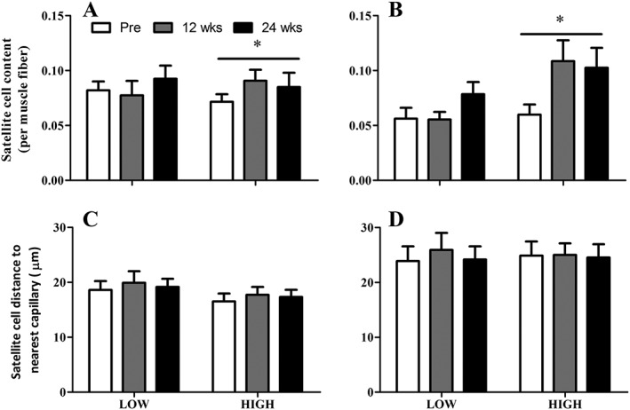
Methods: Twenty-two healthy older men (71 ± 1 years) performed 24 weeks of progressive resistance type exercise training. To assess the change in muscle fibre characteristics, percutaneous biopsies from the vastus lateralis muscle were taken before and following 12 and 24 weeks of the intervention programme. A comparison was made between participants who had a relatively low type II muscle fibre capillary-to-fibre perimeter exchange index (CFPE; LOW group) and high type II muscle fibre CFPE (HIGH group) at baseline. Type I and type II muscle fibre size, satellite cell, capillary content and distance between satellite cells to the nearest capillary were determined by immunohistochemistry.
Results: Overall, type II muscle fibre size (from 5150 ± 234 to 6719 ± 446 µm2 , P < 0.05) and satellite cell content (from 0.058 ± 0.006 to 0.090 ± 0.010 satellite cells per muscle fibre, P < 0.05) had increased significantly in response to 24 weeks of resistance exercise training. However, these improvements where mainly driven by differences in baseline type II muscle fibre capillarization, whereas muscle fibre size (from 5170 ± 390 to 7133 ± 314 µm2 , P < 0.05) and satellite cell content (from 0.059 ± 0.009 to 0.102 ± 0.017 satellite cells per muscle fibre, P < 0.05) increased significantly in the HIGH group, no significant changes were observed in LOW group following exercise training. No significant changes in type I and type II muscle fibre capillarization were observed in response to 12 and 24 weeks of resistance exercise training in both the LOW and HIGH group.
Conclusions: Type II muscle fibre capillarization at baseline may be a critical factor for allowing muscle fibre hypertrophy to occur during prolonged resistance exercise training in older men.
Keywords: Capillary; Elderly; Exercise training; Fibre growth; Skeletal muscle.
© 2016 The Authors. Journal of Cachexia, Sarcopenia and Muscle published by John Wiley & Sons Ltd on behalf of the Society on Sarcopenia, Cachexia and Wasting Disorders.
Figures


References
-
- Cruz‐Jentoft AJ, Baeyens JP, Bauer JM, Boirie Y, Cederholm T, Landi F, et al. Sarcopenia: European consensus on definition and diagnosis: report of the European working group on sarcopenia in older people. Age Ageing 2010;39:412–423, doi:10.1093/ageing/afq034. - DOI - PMC - PubMed
-
- Clynes MA, Edwards MH, Buehring B, Dennison EM, Binkley N, Cooper C. Definitions of sarcopenia: associations with previous falls and fracture in a population sample. Calcif Tissue Int 2015;97:445–452, doi:10.1007/s00223-015-0044-z. - DOI - PMC - PubMed
-
- Janssen I, Baumgartner RN, Ross R, Rosenberg IH, Roubenoff R. Skeletal muscle cutpoints associated with elevated physical disability risk in older men and women. Am J Epidemiol 2004;159:413–421. - PubMed
-
- Janssen I, Heymsfield SB, Ross R. Low relative skeletal muscle mass (sarcopenia) in older persons is associated with functional impairment and physical disability. J Am Geriatr Soc 2002;50:889–896. - PubMed
-
- Landi F, Liperoti R, Russo A, Giovannini S, Tosato M, Capoluongo E, et al. Sarcopenia as a risk factor for falls in elderly individuals: results from the ilSIRENTE study. Clin Nutr 2012;31:652–658, doi:10.1016/j.clnu.2012.02.007. - DOI - PubMed
MeSH terms
LinkOut - more resources
Full Text Sources
Other Literature Sources
