. 2016 Nov 26;17(12):1979.
doi: 10.3390/ijms17121979.

The Antiproliferative Effect of Chakasaponins I and II, Floratheasaponin A, and Epigallocatechin 3-O-Gallate Isolated from Camellia sinensis on Human Digestive Tract Carcinoma Cell Lines

Affiliations

The Antiproliferative Effect of Chakasaponins I and II, Floratheasaponin A, and Epigallocatechin 3-O-Gallate Isolated from Camellia sinensis on Human Digestive Tract Carcinoma Cell Lines

Niichiro Kitagawa et al. Int J Mol Sci. .

Abstract

Acylated oleanane-type triterpene saponins, namely chakasaponins I (1) and II (2), floratheasaponin A (3), and their analogs, together with catechins-including (-)-epigallocatechin 3-O-gallate (4), flavonoids, and caffeine-have been isolated as characteristic functional constituents from the extracts of "tea flower", the flower buds of Camellia sinensis (Theaceae), which have common components with that of the leaf part. These isolates exhibited antiproliferative activities against human digestive tract carcinoma HSC-2, HSC-4, MKN-45, and Caco-2 cells. The antiproliferative activities of the saponins (1-3, IC50 = 4.4-14.1, 6.2-18.2, 4.5-17.3, and 19.3-40.6 µM, respectively) were more potent than those of catechins, flavonoids, and caffeine. To characterize the mechanisms of action of principal saponin constituents 1-3, a flow cytometric analysis using annexin-V/7-aminoactinomycin D (7-AAD) double staining in HSC-2 cells was performed. The percentage of apoptotic cells increased in a concentration-dependent manner. DNA fragmentation and caspase-3/7 activation were also detected after 48 h. These results suggested that antiproliferative activities of 1-3 induce apoptotic cell death via activation of caspase-3/7.

Keywords: (–)-epigallocatechin 3-O-gallate; Camellia sinensis; anti-proliferative activity; apoptosis; chakasaponin; floratheasaponin; tea flower.


Conflict of interest statement

The authors declare no conflict of interest.

Figures

Figure 1
Figure 1
Structures of chakasaponins I (1) and II (2), floratheasaponin A (3), and (–)-epigallocatechin 3-O-gallate (4).
Figure 2
Figure 2
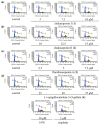
Effects of 14, 5-FU, and cisplatin on the cell cycle distribution of HSC-2 cells. Cell cycle distribution was measured by Muse® Cell Analyzer using a Muse Cell Cycle Kit (Merck Millipore, Darmstadt, Germany); HSC-2 cells were treated with (a) 5, 7.5, and 10 µM of 1; (b) 10, 12.5, and 15 µM of 2; (c) 2.5, 5, and 7.5 µM of 3; (d) 25, 50, and 75 µM of 4, 30 µM of 5-FU, and 3 µM of cisplatin for 48 h; the data represent the mean percentages ± SD of total apoptosis (n = 3); commercial 5-FU and cisplatin were purchased from Wako Pure Chemical Industries, Ltd. (Osaka, Japan).
Figure 3
Figure 3
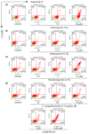
Effects of 14 and camptothecin on the apoptosis of HSC-2 cells. Annexin-V-binding was measured by Muse® Cell Analyzer using a Muse Annexin-V and Deal Cell Kit (Merck Millipore); HSC-2 cells were treated with (a) 5, 7.5, and 10 µM of 1; (b) 10, 12.5, and 15 µM of 2; (c) 2.5, 5, and 7.5 µM of 3; (d) 25, 50, and 75 µM of 4 and 0.02 and 0.05 µM of camptothecin for 24 h; the data represent the mean percentages ± SD of total apoptosis (n = 3); commercial camptothecin was purchased from Wako Pure Chemical Industries, Ltd. (Osaka, Japan).
Figure 4
Figure 4
Morphological analysis of HSC-2 cells treated with 14 and camptothecin. Morphology of representative fields of HSC-2 cells stained with 4′,6-diamidino-2-phenylindole dihydrochloride (DAPI) after treatment with (a) 5, 7.5, and 10 µM of 1; (b) 10, 15, and 17.5 µM of 2; (c) 2.5, 5, and 7.5 µM of 3; (d) 25, 50, and 75 µM of 4 and 0.05 µM of camptothecin for 48 h; the cells indicated by arrows represent fragmented and condensed nuclear chromatins; commercial camptothecin was purchased from Wako Pure Chemical Industries, Ltd. (Osaka, Japan).
Figure 4
Figure 4
Morphological analysis of HSC-2 cells treated with 14 and camptothecin. Morphology of representative fields of HSC-2 cells stained with 4′,6-diamidino-2-phenylindole dihydrochloride (DAPI) after treatment with (a) 5, 7.5, and 10 µM of 1; (b) 10, 15, and 17.5 µM of 2; (c) 2.5, 5, and 7.5 µM of 3; (d) 25, 50, and 75 µM of 4 and 0.05 µM of camptothecin for 48 h; the cells indicated by arrows represent fragmented and condensed nuclear chromatins; commercial camptothecin was purchased from Wako Pure Chemical Industries, Ltd. (Osaka, Japan).
Figure 5
Figure 5
DNA fragmentation in HSC-2 cells treated with 14 and actinomycin D. Representative DNA fragmentation of HSC-2 treated with (a) 5, 10, and 15 µM of 1; (b) 10, 15, and 20 µM of 2; (c) 2.5, 5, and 7.5 µM of 3; (d) 25, 50, and 75 µM of 4, and (Act.D) 0.1 µM of actinomycin D for 48 h; (M) represents a marker (100 bp DNA ladder); commercial actinomycin D was purchased from Wako Pure Chemical Industries, Ltd. (Osaka, Japan).
Figure 6
Figure 6
Effects of 14 and camptothecin on caspase-3/7 in HSC-2 cells. Activities of caspase-3/7 were measured by Muse® Cell Analyzer using a Muse Caspase-3/7 Kit (Merck Millipore); HSC-2 cells were treated with (a) 5, 7.5, and 10 µM of 1; (b) 10, 12.5, and 15 µM of 2; (c) 2.5, 5, and 7.5 µM of 3; (d) 25, 50, and 75 µM of 4 and 0.03 and 0.05 µM of camptothecin for 24 h; the data represent the mean percentages ± SD of total apoptosis (n = 3); commercial camptothecin was purchased from Wako Pure Chemical Industries, Ltd. (Osaka, Japan).

Similar articles

Cited by

References

    1. Sparg S.G., Light M.E., van Staden J. Biological activities and distribution of plant saponins. J. Ethnopharmacol. 2004;94:219–243. doi: 10.1016/j.jep.2004.05.016. - DOI - PubMed
    1. Vincken J.-P., Heng L., de Groot A., Gruppen H. Saponins, classification and occurrence in the plant kingdom. Phytochemistry. 2007;68:275–297. doi: 10.1016/j.phytochem.2006.10.008. - DOI - PubMed
    1. Podolak I., Galanty A., Sobolewska D. Saponins as cytotoxic agents: A review. Phytochem. Rev. 2010;9:425–474. doi: 10.1007/s11101-010-9183-z. - DOI - PMC - PubMed
    1. Dinda B., Debnath S., Mohanta B.C., Harigaya Y. Naturally occurring triterpenoid saponins. Chem. Biodivers. 2010;7:2327–2580. doi: 10.1002/cbdv.200800070. - DOI - PubMed
    1. Osbourn A., Goss R.J.M., Field R.A. The saponins—Polar isoprenoids with important and diverse biological activities. Nat. Prod. Rep. 2011;28:1261–1268. doi: 10.1039/c1np00015b. - DOI - PubMed