

Lamina cribrosa in glaucoma
- PMID: 27898470
- PMCID: PMC5480216
- DOI: 10.1097/ICU.0000000000000354
Lamina cribrosa in glaucoma
Abstract
Purpose of review: This article presents, summarizes, and interprets the most recent advances in the study and understanding of the lamina cribrosa in glaucoma, in the context of previous work.
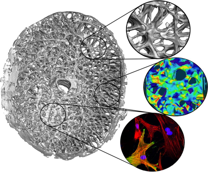
Recent findings: The lamina is an active living structure that responds to strain by changing morphology at the micro-scale and macro-scale in glaucoma. Changes in lamina cribrosa morphology in glaucoma include posteriorization of the laminar insertion into the sclera, increased cupping or depth of the lamina cribrosa, and the development of focal lamina cribrosa defects. These lamina cribrosa changes are associated with disk hemorrhages and visual field damage, and are detectable with clinical imaging techniques such as optical coherence tomography. Glaucomatous changes in the lamina cribrosa are thought to be driven by cellular processes mediated by focal cyclical mechanical strain. Strain is eye specific and mediated by intraocular pressure, cerebrospinal fluid pressure, scleral and lamina cribrosa morphology, and structural stiffness; deleterious lamina cribrosa strains can occur at all levels of mean intraocular pressure.
Summary: Laminar morphology is ever changing in health and disease, and recent studies have identified several promising morphological changes that are indicative of glaucoma susceptibility, onset, and progression.
Conflict of interest statement
The authors have no relevant conflicts of interest
Figures
References
-
- Agoumi Y, et al. Laminar and prelaminar tissue displacement during intraocular pressure elevation in glaucoma patients and healthy controls. Ophthalmology. 2011;118(1):52–9. - PubMed
Publication types
MeSH terms
Grants and funding
LinkOut - more resources
Full Text Sources
Other Literature Sources
Medical
Research Materials
