

Genetic control of postnatal human brain growth
- PMID: 27898583
- PMCID: PMC5340196
- DOI: 10.1097/WCO.0000000000000405
Genetic control of postnatal human brain growth
Abstract
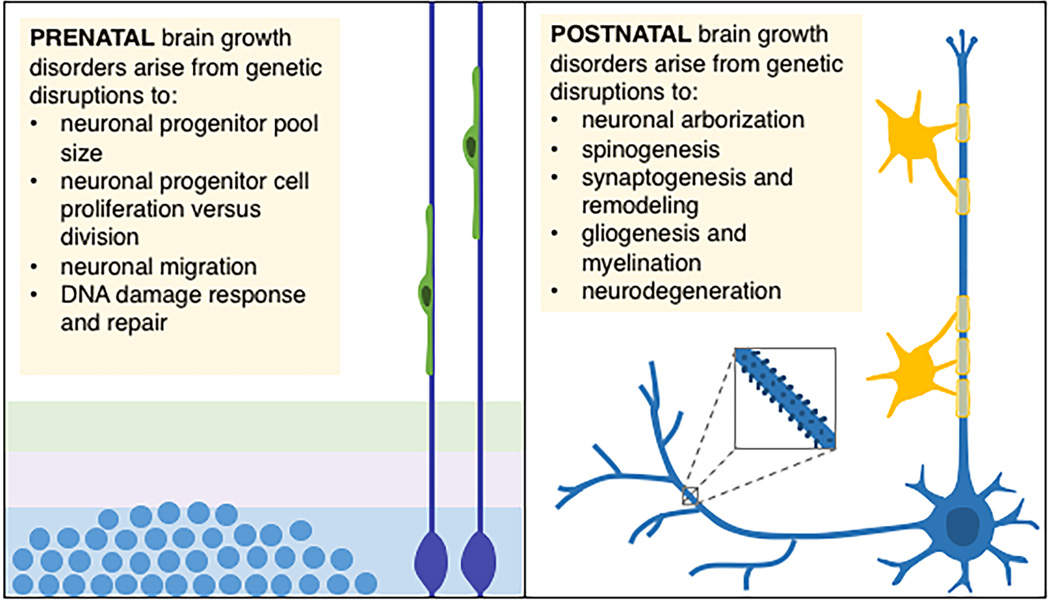
Purpose of review: Studies investigating postnatal brain growth disorders inform the biology underlying the development of human brain circuitry. This research is becoming increasingly important for the diagnosis and treatment of childhood neurodevelopmental disorders, including autism and related disorders. Here, we review recent research on typical and abnormal postnatal brain growth and examine potential biological mechanisms.
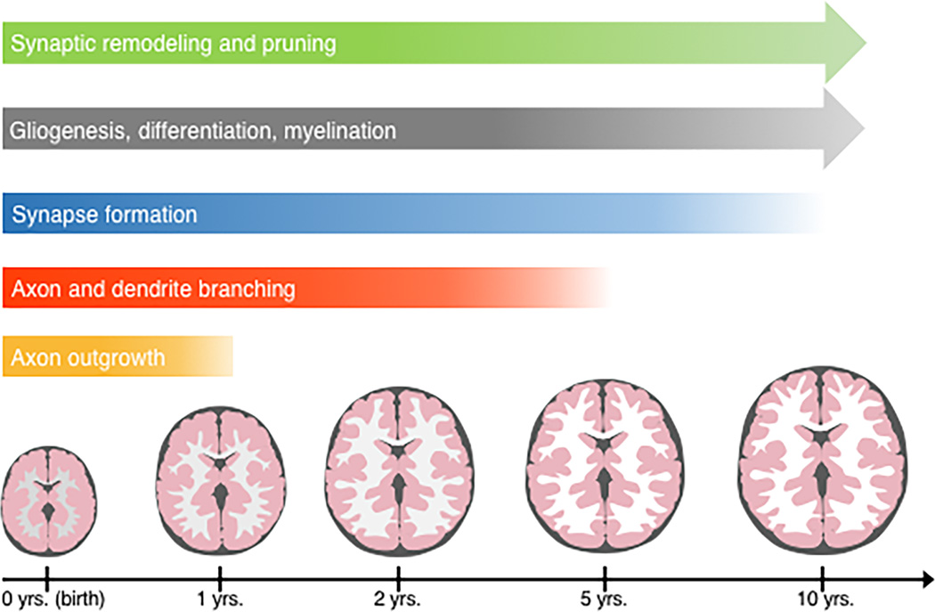
Recent findings: Clinically, brain growth disorders are heralded by diverging head size for a given age and sex, but are more precisely characterized by brain imaging, post-mortem analysis, and animal model studies. Recent neuroimaging and molecular biological studies on postnatal brain growth disorders have broadened our view of both typical and pathological postnatal neurodevelopment. Correlating gene and protein function with brain growth trajectories uncovers postnatal biological mechanisms, including neuronal arborization, synaptogenesis and pruning, and gliogenesis and myelination. Recent investigations of childhood neurodevelopmental and neurodegenerative disorders highlight the underlying genetic programming and experience-dependent remodeling of neural circuitry.
Summary: To understand typical and abnormal postnatal brain development, clinicians and researchers should characterize brain growth trajectories in the context of neurogenetic syndromes. Understanding mechanisms and trajectories of postnatal brain growth will aid in differentiating, diagnosing, and potentially treating neurodevelopmental disorders.
Conflict of interest statement
None
Figures
References
-
- Rollins JD, Collins JS, Holden KR. United States head circumference growth reference charts: birth to 21 years. J Pediatr. 2010;156:907–913. - PubMed
-
- Kuczmarski RJ, Ogden CL, Guo SS, et al. 2000 CDC Growth Charts for the United States: methods and development. Vital Health Stat. 2002;11:1–190. - PubMed
-
- Bartholomeusz HH, Courchesne E, Karns CM. Relationship between head circumference and brain volume in healthy normal toddlers, children, and adults. Neuropediatrics. 2002;33:239–241. - PubMed
-
- Lindley AA, Benson JE, Grimes C, et al. The relationship in neonates between clinically measured head circumference and brain volume estimated from head CT-scans. Early Hum Dev. 1999;56:17–29. - PubMed
Publication types
MeSH terms
Grants and funding
LinkOut - more resources
Full Text Sources
Other Literature Sources
Medical
Research Materials
