. 2017 Jan 17;116(2):218-226.
doi: 10.1038/bjc.2016.394. Epub 2016 Nov 29.

Overexpression of PBK/TOPK relates to tumour malignant potential and poor outcome of gastric carcinoma

Affiliations

Overexpression of PBK/TOPK relates to tumour malignant potential and poor outcome of gastric carcinoma

Takuma Ohashi et al. Br J Cancer. .

Abstract

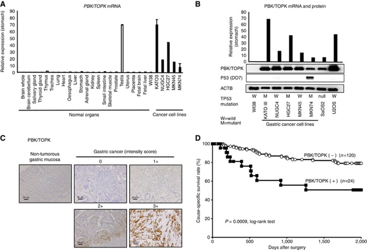
Background: PDZ-binding kinase/T-LAK cell-originated protein kinase (PBK/TOPK) is a serine-threonine kinase and overexpressed in various types of cancer by inhibiting the transactivation activities of p53 and PTEN. We tested whether PBK/TOPK acts as a cancer-promoting gene through its activation/overexpression in gastric cancer (GC).

Methods: We analysed five GC cell lines and 144 primary tumours, which were curatively resected in our hospital between 2001 and 2003.

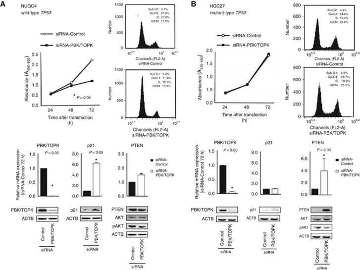
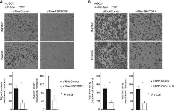
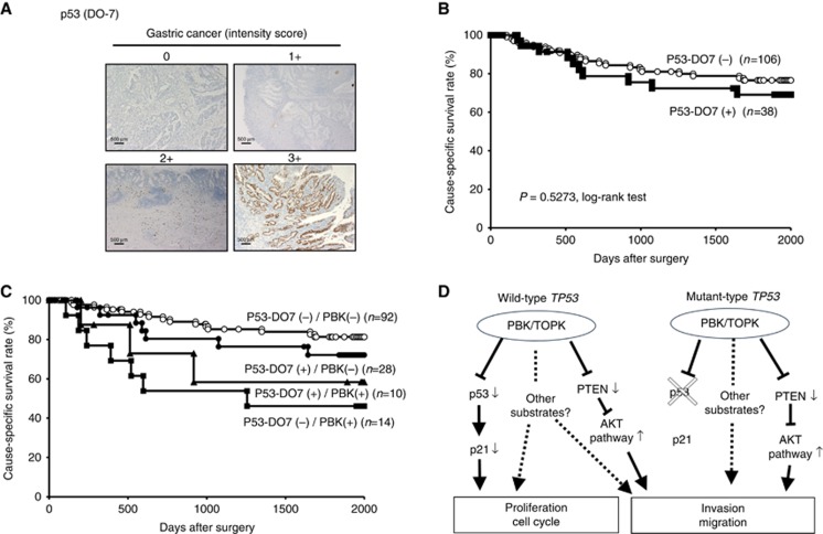
Results: Overexpression of the PBK/TOPK protein was frequently detected in GC cell lines (4 out of 5 lines, 80.0%) was detected in primary tumour samples of GC (24 out of 144 cases, 16.6%) and was significantly correlated with venous invasion, tumour depth and recurrence rate. PDZ-binding kinase/T-LAK cell-originated protein kinase-overexpressing tumours had a worse survival rate than those with non-expressing tumours (P=0.0009, log-rank test). PDZ-binding kinase/T-LAK cell-originated protein kinase positivity was independently associated with a worse outcome in multivariate analysis (P<0.0001, hazard ratio 6.40 (2.71-14.49)). In PBK/TOPK-overexpressing GC cells, knockdown of PBK/TOPK inhibited the cell proliferation through the p53 activation in a TP53 mutation-dependent manner and inhibited the migration/invasion through the PTEN upregulation in a TP53 mutation-independent manner.

Conclusions: These findings suggest PBK/TOPK plays a crucial role in tumour malignant potential through its overexpression and highlight its usefulness as a prognostic factor and potential therapeutic target in GC.


Figures

Figure 1
Figure 1
Expression of PBK/TOPK was correlated with poor prognosis in gastric cancer patients. (A) Expression of PBK/TOPK mRNA in five GC cell lines compared with that of the normal organs and the fibroblast cell line WI-38. (B) Expression of PBK/TOPK mRNA and protein in five GC cell lines and osteosarcoma cell lines, such as TP53-null SaOS2 and TP53 wild-type U2OS, compared with that in the fibroblast cell line WI-38. The status of TP53 mutation in each cancer cell line was evaluated by western blotting. The status of TP53 mutation was positively associated with the reported status of TP53 mutation in the database (http://p53.free.fr/index.html, W: wild-type TP53, M: mutant TP53). (C) Specific immunostaining of PBK/TOPK in a representative primary tumour sample. On the basis of this result, the intensity scores for PBK/TOPK staining were determined as follows: 0=negative, 1=weak, 2=moderate, 3=strong. Magnification: × 200; Scale bar, 20 μm. (D) Cause-specific survival rates of GC patients (as determined by Kaplan–Meier plots) depending on the combination scores of intensity and proportion in PBK/TOPK expression.
Figure 2
Figure 2
Effects of PBK/TOPK knockdown by siRNA-PBK/TOPK compared with those of control siRNA in NUGC4 and HGC27 cell lines. (A) shows that the knockdown of PBK/TOPK overexpression in TP53 wild-type NUGC4 cells downregulated expression of pAKT and upregulated that of p21, which resulted mainly in G0–G1 arrest. The proliferation of NUGC4 cell was significantly lower than with controls after the knockdown of endogenous PBK/TOPK expression. However, (B) shows that there was no difference of cell proliferation and FACS results between siRNA-PBK/TOPK and control siRNA cells in TP53 mutant HGC27 cells. The p21 level was not changed, but pAKT level was decreased and PTEN level was increased at 72 h in siRNA-PBK/TOPK-transfected cells. Including the results of Supplementary Figure S2, these results suggested that PBK/TOPK might be related to the cell proliferation in a TP53 mutation-dependent manner.
Figure 3
Figure 3
Regarding the cell migration and invasion, the results of the increase of PTEN and/or the decrease of pAKT in siRNA-PBK/TOPK-transfected cells suggested that PBK/TOPK may increase the ability of GC cells to migrate and invade via the PI3K/Akt pathway activation in a TP53 mutation-independent manner. (A) NUGC wild-type TP53 and (B) mutant-type TP53.
Figure 4
Figure 4
Relationship between protein expression of PBK/TOPK and p53 in primary cases of gastric cancer. (A) Specific immunostaining of PBK/TOPK in a representative primary tumour sample. On the basis of this result, the intensity scores for p53 (DO7) staining were determined as follows: 0=negative, 1=weak, 2=moderate, 3=strong. Magnification: × 40; Scale bar, 500 μm. (B) Kaplan–Meier curves for cause-specific survival rates of patients at all stages according to the expression of p53. There was no difference of survival between the patients positive and negative for p53-DO7. (C) Postoperative overall survival curves according to a combination of the expression of PBK/TOPK and p53. A significant difference in survival was observed among the patients positive (+) or negative (−) for p53-DO7 and PBK/TOPK (P=0.0228, log-rank test). Notably, in the p53-DO7-negative group, the patients with PBK/TOPK expression had a markedly worse outcome than those with no PBK/TOPK expression. (D) Hypothetical model of the overexpression/activation of PBK/TOPK in GC cells.

References

    1. Abe Y, Matsumoto S, Kito K, Ueda N (2000) Cloning and expression of a novel MAPKK-like protein kinase, lymphokine-activated killer T-cell-originated protein kinase, specifically expressed in the testis and activated lymphoid cells. J Biol Chem 275(28): 21525–21531. - PubMed
    1. Ayllon V, O'Connor R (2007) PBK/TOPK promotes tumour cell proliferation through p38 MAPK activity and regulation of the DNA damage response. Oncogene 26(24): 3451–3461. - PubMed
    1. Bang YJ, Van Cutsem E, Feyereislova A, Chung HC, Shen L, Sawaki A, Lordick F, Ohtsu A, Omuro Y, Satoh T, Aprile G, Kulikov E, Hill J, Lehle M, Ruschoff J, Kang YK (2010) Trastuzumab in combination with chemotherapy versus chemotherapy alone for treatment of HER2-positive advanced gastric or gastro-oesophageal junction cancer (ToGA): a phase 3, open-label, randomised controlled trial. Lancet 376(9742): 687–697. - PubMed
    1. Becker KF, Atkinson MJ, Reich U, Becker I, Nekarda H, Siewert JR, Hofler H (1994) E-cadherin gene mutations provide clues to diffuse type gastric carcinomas. Cancer Res 54(14): 3845–3852. - PubMed
    1. Bellini MF, Cadamuro AC, Succi M, Proenca MA, Silva AE (2012) Alterations of the TP53 gene in gastric and esophageal carcinogenesis. J Biomed Biotechnol 2012: 891961. - PMC - PubMed

Substances