. 2016 Nov 29;12(11):e1006321.
doi: 10.1371/journal.pgen.1006321. eCollection 2016 Nov.

High-Resolution Phenotypic Landscape of the RNA Polymerase II Trigger Loop

Affiliations

High-Resolution Phenotypic Landscape of the RNA Polymerase II Trigger Loop

Chenxi Qiu et al. PLoS Genet. .

Erratum in

Abstract

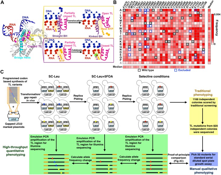
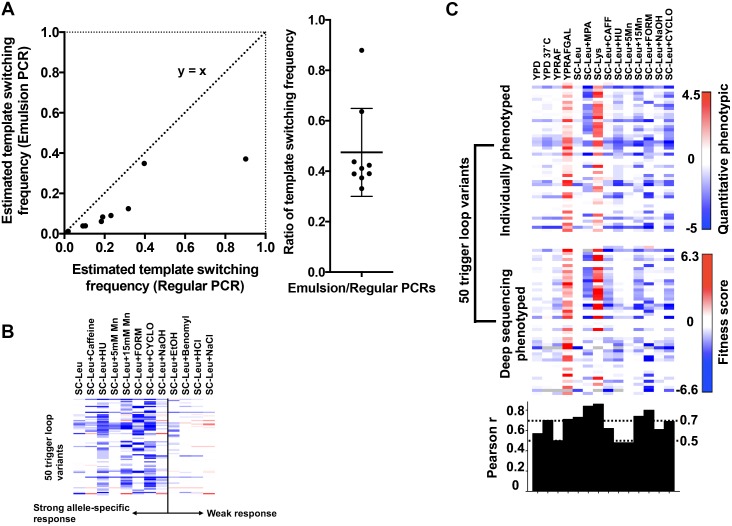
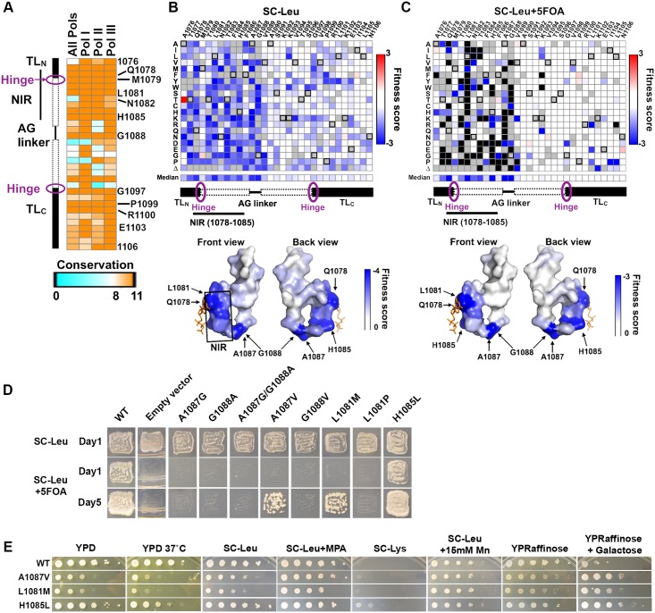
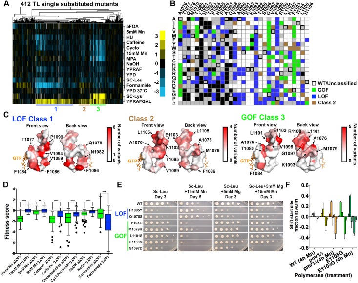
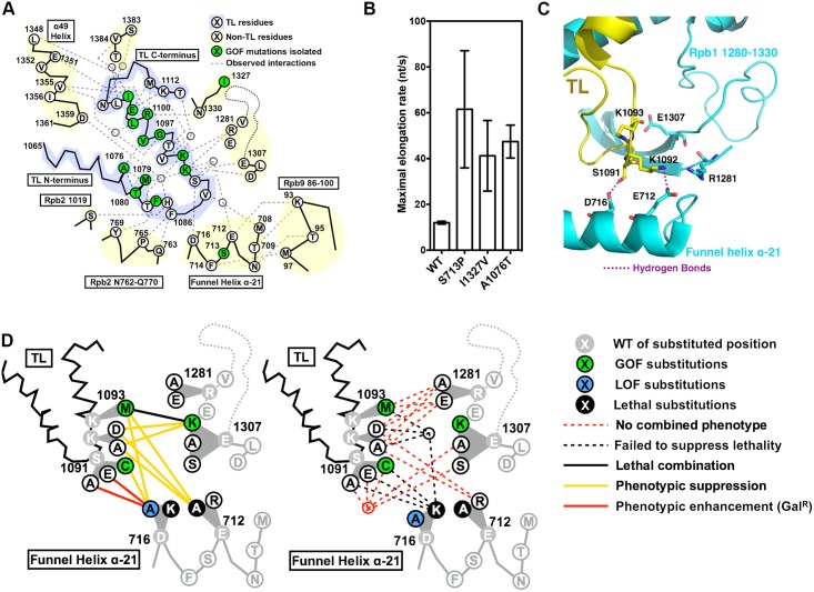
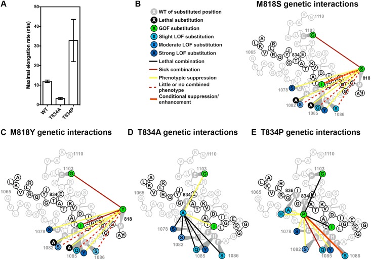
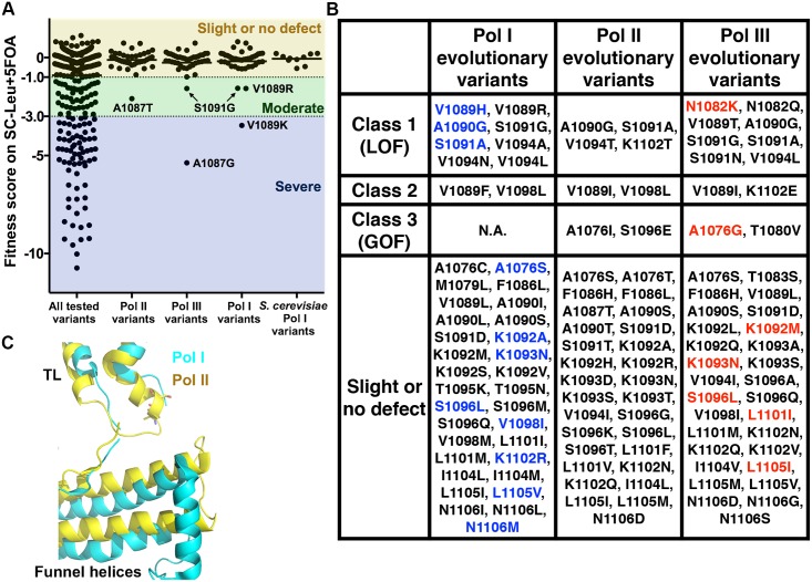
The active sites of multisubunit RNA polymerases have a "trigger loop" (TL) that multitasks in substrate selection, catalysis, and translocation. To dissect the Saccharomyces cerevisiae RNA polymerase II TL at individual-residue resolution, we quantitatively phenotyped nearly all TL single variants en masse. Three mutant classes, revealed by phenotypes linked to transcription defects or various stresses, have distinct distributions among TL residues. We find that mutations disrupting an intra-TL hydrophobic pocket, proposed to provide a mechanism for substrate-triggered TL folding through destabilization of a catalytically inactive TL state, confer phenotypes consistent with pocket disruption and increased catalysis. Furthermore, allele-specific genetic interactions among TL and TL-proximal domain residues support the contribution of the funnel and bridge helices (BH) to TL dynamics. Our structural genetics approach incorporates structural and phenotypic data for high-resolution dissection of transcription mechanisms and their evolution, and is readily applicable to other essential yeast proteins.


Conflict of interest statement

Authors JVdB and RS are employed by MorphoSys AG, which currently holds rights to the Slonomics DNA synthesis technology utilized in the manuscript.

Figures

Fig 1
Fig 1. Establishment of a high-throughput platform for phenotyping comprehensive TL single variant library.
(A) Multiple TL functions are underpinned by its mobile nature. Structures of open (PDB:5C4J) and closed TLs (PDB:2E2H) are shown in the context of surrounding domains. Template DNA (blue), RNA (red), Bridge Helix (cyan), Closed TL (magenta) and Open TL (yellow) are shown in cartoon rendering. The open TL has been proposed to allow Pol II translocation while the closed TL has been shown to facilitate catalysis (right panel). (B) Mutational coverage of the TL variant library is shown as a heatmap illustrating the allele frequencies of single substitution variants (WT amino acids and positions labeled on x axis; amino acid substitutions on y axis). The WT amino acids for each position are noted with black boxes, and mutants excluded from library synthesis are noted using blue boxes. (C) Schematic representation of experimental approach. Stars of different colors represent distinct substitutions. The TL variant library PCR amplicon (encoding Rpb1 amino acids 1076–1106) flanked by RPB1 TL flanking sequence (orange) was co-transformed with a linearized LEU2 CEN plasmid containing an rpb1 gene with the TL deleted, allowing construction of full-length RPB1 (with TL variants) by in vivo homologous recombination. Heterozygous Leu+ transformants were replica-plated onto SC-Leu+5FOA to select against the WT RPB1 (URA3 CEN) plasmid and to create TL variant pools. TL variant pools were subsequently replica-plated to different selective conditions for either traditional individual colony screening or high-throughput phenotyping using deep sequencing. For the latter, replica-plated colonies were pooled for genomic DNA extraction, and the TL region was amplified by emulsion PCR to prepare templates for deep sequencing.
Fig 2
Fig 2. Quality controls for the TL high-throughput phenotyping approach.
(A) Comparison of estimated template switching frequencies in regular and emulsion PCR conditions. Template switching was estimated by the ratio (FreqDouble) / (FreqSingle1 × FreqSingle2) for all the possible double mutants combined from five spiked-in single mutants. (B) Additional growth conditions were employed to increase resolution for distinguishing similar TL alleles. Growth scores for 50 individually isolated TL mutants (y axis) under 12 growth conditions (x axis), as determined by standard serial dilution plate phenotyping (S2 and S3 Figs), are shown as a heatmap. Positive values shown in red indicate increase in allele frequency relative to WT and negative values in blue indicate decrease in allele frequency relative to WT. (C) High-throughput quantitative phenotyping results are consistent with individual phenotyping of variants. Top heatmap shows qualitative growth scores (as in Fig 2B) of 50 individually phenotyped TL variants on the y axis (S2–S4 Figs) with selective conditions on the x axis. Deep sequencing results for the same mutants using median of fitness defects from three independent high-throughput screens are shown in the middle panel. Pearson r calculated to show the correlation between each condition from the two datasets is shown in the bottom panel.
Fig 3
Fig 3. The TL fitness landscape distinguishes highly conserved TL residues and reveals high mutational sensitivity in the nucleotide interacting region (NIR) and the Alanine-Glycine linker.
(A) Conservation heatmap of TL residues in eukaryotic RNA polymerases. The conservation scores were extracted from a multiple sequence alignment, including 182 Pol II, 59 Pol I, and 111 Pol III sequences utilizing the conservation metric from Jalview 2.8 version 14.0 [91]. (B) Fitness defects of TL variants in the heterozygous state are shown as a heatmap. Unavailable data points are denoted by filled grey squares. WT residues at indicated positions are denoted by black boxes. Surface representation (bottom panel) of the TL structure (PDB:2E2H) is shaded by the median fitness value for all available variants at each position, in a gradient of white (rare defects) to blue (common defects). The position of the matched GTP substrate is shown in orange stick representation. (C) General fitness defects of TL variants upon removal of WT RPB1. Fitness defects predicted to result in lethality shown in black. Surface representation (bottom panel) of the TL structure is shaded by the median fitness value of all available variants at each position, in a gradient of white (rare defects) to blue (common defects). (D) Complementation abilities of variants in the difficult-to-substitute TL positions (L1081, A1087, G1088) or unexpected TL variants (H1085L) assayed by plasmid shuffling of individual strains. Ability to grow on SC-Leu+5FOA indicates complementation of essential functions of RPB1. SC-Leu medium is the control state where WT RPB1 is present. (E) Transcription-linked phenotypes of viable substitutions in difficult-to-substitute residues (L1081M, A1087V) or a TL variant with unexpectedly mild fitness defects (H1085L).
Fig 4
Fig 4. Three distinct TL mutant classes, revealed from TL phenotypic landscape, have specific distribution on the TL structure and distinct stress response profiles.
(A) Hierarchical clustering of 412 single TL variants’ (x axis) phenotypes (calculated as in Fig 1C) under 14 different conditions (y axis) reveals distinct mutant classes. Positive (yellow) and negative (blue) fitness scores are shown as a heatmap. Mutant classes (clusters) are annotated by colored lines beneath the heatmap. (B) Distribution of three major mutant classes is shown in a single substitution variant heatmap. Class 1 (genetic GOF) mutants are shown in green; Class 2 mutants are shown in brown and Class 3 (genetic LOF) mutants are shown in blue. (C) Distribution of different mutant classes on the TL structure. TL is shown in surface and colored in the gradient from white to red by the number of clustered mutants at each position. (D) Differential stress responses in genetic GOF and LOF mutants. Genetic GOF mutants are more sensitive to Mn2+, caffeine and cycloheximide, whereas genetic LOF mutants are more sensitive to formamide. **p<0.01, ****p<0.0001 (Two-tailed unpaired t-test). (E) Differential Mn2+ sensitivity and its suppression by Mg2+ for selected TL variants representative of mutant classes. (F) Mn2+ effects on different mutants’ transcription start sites (TSSs) distribution at ADH1, determined by primer extension analysis. TSSs at ADH1 are distributed in a range of positions and were divided into six bins for quantitation: from upstream (left) to downstream (right). Change of TSSs (normalized to untreated WT) is calculated by the change in TSS fraction for each bin relative to the WT distribution. Average and standard deviation of three experimental replicates are shown as a bar graph with error bars.
Fig 5
Fig 5. Functional contribution of TL tip and Funnel Helix α-21 to proper TL dynamics.
(A) Observed and predicted interactions between TL and TL-proximal domains. TL schematic is shown with residues identified by single-letter amino acid code and positions of interest annotated. Positions of GOF mutants isolated in our screen, along with the positions for a subset of previously isolated TL-proximal GOF mutants, are color coded in green. Observed TL interactions with other Rpb1 domains from structures or simulation studies are shown as grey dashed lines. (B) Maximal in vitro elongation rates (nucleotides/second) of Pol II WT and genetic GOF mutants S713P, I1327V and A1076T. (C) Observed interactions between open TL tip and TL adjacent charged residues (PDB: 5C4X). Funnel Helix refers to the Rpb1 α-21 alpha-helix. (D) Genetic interactions between the TL tip and proximal Rpb1 domains. Schematics of the TL and adjacent domains are shown in lines, with positions of interest shown in single-letter amino acid code. Substituted residues are shown in grey, with substituting amino acids shown in white, blue or green filled circles based on single substitution phenotypes (S8F Fig). Double substitution phenotypes are shown as colored lines connecting the two relevant single substitutions. Some sets of similar interactions were grouped into nodes to reduce complexity in interaction lines.
Fig 6
Fig 6. Functional interplay between the TL and Bridge Helix (BH).
(A) Maximal in vitro elongation rates (nucleotides/second) of BH variants T834A and T834P. (B) Genetic interactions between BH M818S and TL substitutions. M818S suppressed (yellow lines) the strong LOF TL variants (dark blue) but not the slight and moderate LOF TL variants (light blue), and showed synthetic sickness (red lines) with the GOF TL variants (green). (C) Genetic interactions between BH M818Y and TL substitutions. Similar to M818S genetic interactions with TL variants (Fig 6B), M818Y suppressed (yellow lines) the strong LOF TL variants (dark blue) but not the slight and moderate LOF TL variants (light blue), and showed synthetic sickness (red lines) with GOF TL variants (green). (D) Genetic interactions between BH T834A and TL substitutions. T834A suppressed (yellow lines) the GOF TL variants and was synthetic lethal with all the tested LOF TL variants (blue). (E) Genetic interactions between BH T834P and TL or BH. Similar to M818 variants (Fig 6B, 6C), T834P suppressed strong and moderate LOF TL variants (dark blue) but was synthetic sick with weak LOF TL variants (light blue), while synthetically lethal with GOF TL variants (green). T834P was also suppressed (yellow line) by two LOF BH mutants Y836A/H.
Fig 7
Fig 7. Phenotypic analyses of evolutionary variants suggest context-dependent functions for many TL residues.
(A) General growth fitness defects of the TL single-substituted variants observed in the TL across Pol I, II, III evolution including 38 Pol II, 42 Pol I and 42 Pol III amino acid variants relative to Sce Pol II. (B) Evolutionary TL variants in three mutant classes from the TL phenotypic landscape (Fig 4A and 4B). Existing variants from Sce Pol I are colored in blue, and existing variants from Sce Pol III are colored in red. Sce Pol I has three substitutions (V1089H, A1090G and S1091A) that cause LOF in the Pol II context; Sce Pol III has one substitution (A1076G) classified as GOF and one substitution (N1082K) classified as LOF. (C) Difference in positioning of funnel helices (relative to TL) in Pol I and Pol II. Cartoon representation of TL/funnel helices from Pol I and Pol II are shown in cyan and yellow, respectively (PDB: 5C4J and 2VUM).

References

    1. Gnatt AL, Cramer P, Fu J, Bushnell DA, Kornberg RD (2001) Structural basis of transcription: an RNA polymerase II elongation complex at 3.3 A resolution. Science 292: 1876–1882. 10.1126/science.1059495 - DOI - PubMed
    1. Cramer P, Bushnell DA, Kornberg RD (2001) Structural basis of transcription: RNA polymerase II at 2.8 angstrom resolution. Science 292: 1863–1876. 10.1126/science.1059493 - DOI - PubMed
    1. Westover KD, Bushnell DA, Kornberg RD (2004) Structural basis of transcription: nucleotide selection by rotation in the RNA polymerase II active center. Cell 119: 481–489. 10.1016/j.cell.2004.10.016 - DOI - PubMed
    1. Wang D, Bushnell DA, Westover KD, Kaplan CD, Kornberg RD (2006) Structural basis of transcription: role of the trigger loop in substrate specificity and catalysis. Cell 127: 941–954. 10.1016/j.cell.2006.11.023 - DOI - PMC - PubMed
    1. Liu X, Bushnell DA, Kornberg RD (2013) RNA polymerase II transcription: structure and mechanism. Biochim Biophys Acta 1829: 2–8. 10.1016/j.bbagrm.2012.09.003 - DOI - PMC - PubMed

LinkOut - more resources