. 2016 Nov 29;11(11):e0166784.
doi: 10.1371/journal.pone.0166784. eCollection 2016.

Isoflurane Exposure Induces Cell Death, Microglial Activation and Modifies the Expression of Genes Supporting Neurodevelopment and Cognitive Function in the Male Newborn Piglet Brain

Affiliations

Isoflurane Exposure Induces Cell Death, Microglial Activation and Modifies the Expression of Genes Supporting Neurodevelopment and Cognitive Function in the Male Newborn Piglet Brain

Kevin D Broad et al. PLoS One. .

Abstract

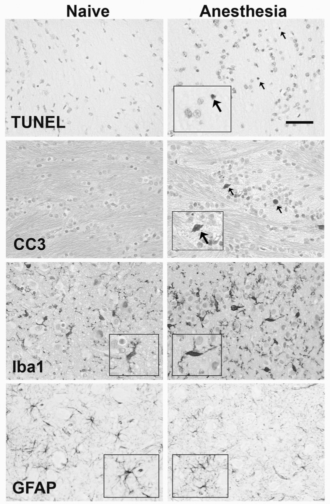
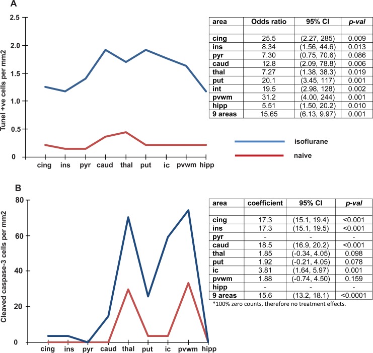
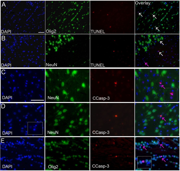
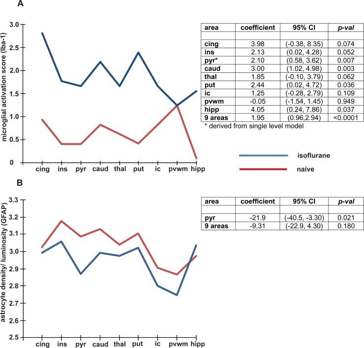
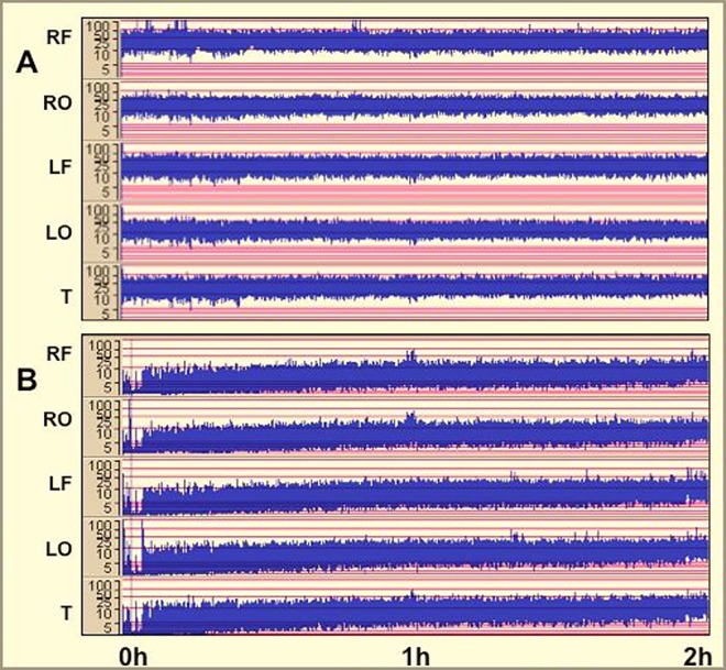
Exposure of the brain to general anesthesia during early infancy may adversely affect its neural and cognitive development. The mechanisms mediating this are complex, incompletely understood and may be sexually dimorphic, but include developmentally inappropriate apoptosis, inflammation and a disruption to cognitively salient gene expression. We investigated the effects of a 6h isoflurane exposure on cell death, microglial activation and gene expression in the male neonatal piglet brain. Piglets (n = 6) were randomised to: (i) naive controls or (ii) 6h isoflurane. Cell death (TUNEL and caspase-3) and microglial activation were recorded in 7 brain regions. Changes in gene expression (microarray and qPCR) were assessed in the cingulate cortex. Electroencephalography (EEG) was recorded throughout. Isoflurane anesthesia induced significant increases in cell death in the cingulate and insular cortices, caudate nucleus, thalamus, putamen, internal capsule, periventricular white matter and hippocampus. Dying cells included both neurons and oligodendrocytes. Significantly, microglial activation was observed in the insula, pyriform, hippocampus, internal capsule, caudate and thalamus. Isoflurane induced significant disruption to the expression of 79 gene transcripts, of these 26 are important for the control of transcription and 23 are important for the mediation of neural plasticity, memory formation and recall. Our observations confirm that isoflurane increases apoptosis and inflammatory responses in the neonatal piglet brain but also suggests novel additional mechanisms by which isoflurane may induce adverse neural and cognitive development by disrupting the expression of genes mediating activity dependent development of neural circuits, the predictive adaptive responses of the brain, memory formation and recall.


Conflict of interest statement

The authors have declared that no competing interests exist.

Figures

Fig 1
Fig 1. Histology of TUNEL, CCasp-3, Iba-1 and GFAP.
Representative photomicrograpths of TUNEL and CCasp-3 in the external capsule and the Iba-1 and GFAP in the thalamus of naïve (left column) and isoflurane exposed (right column) piglets. Arrows indicate TUNEL and CCasp-3 positive cells. CCasp-3 and Iba-1 are counterstained with cresyl violet. Inserts highlight cell activation and morphology (scale bar = 50μm).
Fig 2
Fig 2
Effect of a six hour exposure of isoflurane on (A) TUNEL positive cells, (B) Cleaved caspase 3. The figures on the left show the values relative to brain region in the isoflurane and naïve groups. The tables on the right show the odds ratio (95% CI) and p values for the comparisons between isoflurane exposure and naïve brain in all brain regions as well as the total for the combined 9 regions. Abbreviations; cing = cingulate cortex, ins = insular cortex, pyr = pyriform cortex, caud = caudate nucleus, thal = thalamus, put = putamen, ic = internal capsule, pvwm = periventricular white matter, hipp = hippocampus.
Fig 3
Fig 3. Co-localisation of cell death and lineage markers.
TUNEL and CCasp-3 (red), oligodendrocyte cell lineage marker (Olig-2, green), neuronal cell linage marker (NeuN, green), nuclear stain (DAPI, blue). In the (A) periventricular white matter, (B) dorsoparietal cortex layer 1/2, (C) thalamus, (D) sensoriomotor cortex layer 3/4, (E) external capsule. Cells expressing TUNEL and either Olig-2 or NeuN are indicated with white arrows in the overlay panel (A and B), and those not co-labelling (due to loss of cell specific markers induced by the stage of death or alternate cell type) are indicated with a pink arrow (B, C, D). Scale bar in (A-B) is 50μm and in (C-E) is 25μm.
Fig 4
Fig 4
Effect of a six hour exposure of isoflurane on (A) Microglial activation and (B) Astrogliosis. The figures on the left show the values relative to brain region in the isoflurane and naïve groups. The tables on the right show the odds ratio (95% CI) and p values for the comparisons between isoflurane exposure and naïve brain in all brain regions as well as the total for the combined 9 regions. Abbreviations; cing = cingulate cortex, ins = insular cortex, pyr = pyriform cortex, caud = caudate nucleus, thal = thalamus, put = putamen, ic = internal capsule, pvwm = periventricular white matter, hipp = hippocampus.
Fig 5
Fig 5. Representative amplitude-integrated electroencephalographs (aEEGs) in response to 2h of isoflurane exposure (0 to 2h).
A. Continuous normal voltage (CNV) throughout. B. Low voltage, recovering to CNV by 2h.

References

    1. Brambrink AM, Evers AS, Avidan MS, Farber NB, Smith DJ, Zhang X. et al. Isoflurane-induced neuroapoptosis in the neonatal rhesus macaque brain. Anesthesiology. 2010; 113 (5): 834–41. - PMC - PubMed
    1. Brambrink AM, Back SA, Riddle A, Gong X, Moravec MD, Dissen GA, et al. Isoflurane-induced apoptosis of oligodendrocytes in the neonatal primate brain. Ann Neurol. October 2012; 72 (4): 525–35. 10.1002/ana.23652 - DOI - PMC - PubMed
    1. Creeley CE, Dikranian KT, Dissen GA, Back SA, Olney JW, Brambrink AM. Isoflurane-induced apoptosis of neurons and oligodendrocytes in the fetal rhesus macaque brain. Anesthesiology. 2014. March; 120 (3): 626–38. 10.1097/ALN.0000000000000037 - DOI - PMC - PubMed
    1. Lu LX, Yon J, Carter L, Jevtovic-Todorovic, V. General anesthesia activates BDNF-dependent neuroapoptosis in the developing rat brain. Apoptosis. 2006; 11 (9): 1603–15. 10.1007/s10495-006-8762-3 - DOI - PubMed
    1. Schubert H, Eiselt M, Walter B, Fritz H, Brodhun M, Bauer R . Isoflurane/nitrous oxide anesthesia and stress-induced procedures enhance neuroapoptosis in intrauterine growth-restricted piglets. Intensive Care Med. 2012. July; 38 (7): 1205–14. 10.1007/s00134-012-2576-2 - DOI - PubMed

MeSH terms