. 2016 Nov 29;11(11):e0167250.
doi: 10.1371/journal.pone.0167250. eCollection 2016.

Relationships between Mucosal Antibodies, Non-Typeable Haemophilus influenzae (NTHi) Infection and Airway Inflammation in COPD

Affiliations

Relationships between Mucosal Antibodies, Non-Typeable Haemophilus influenzae (NTHi) Infection and Airway Inflammation in COPD

Karl J Staples et al. PLoS One. .

Erratum in

Abstract

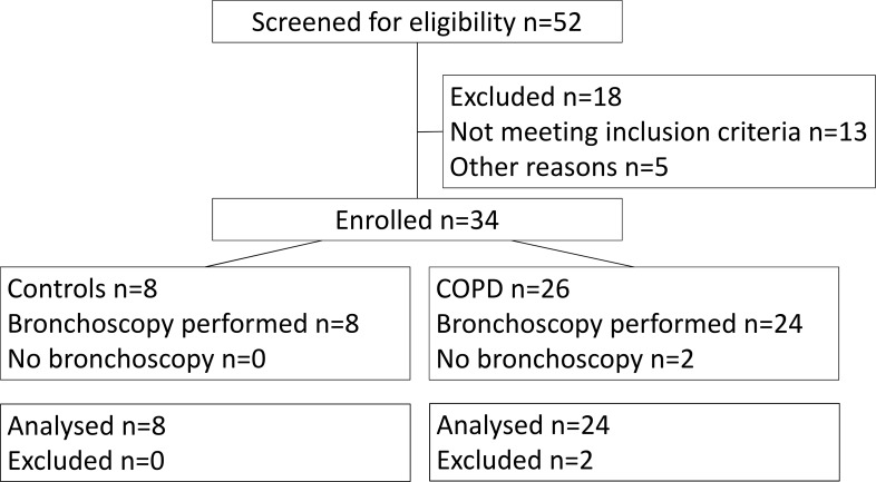
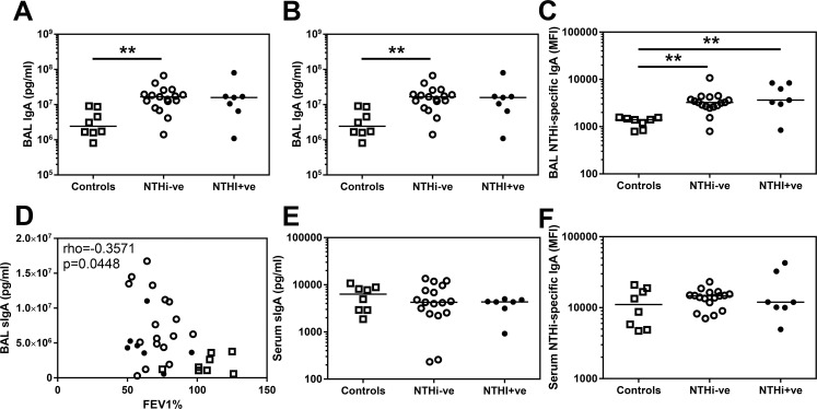
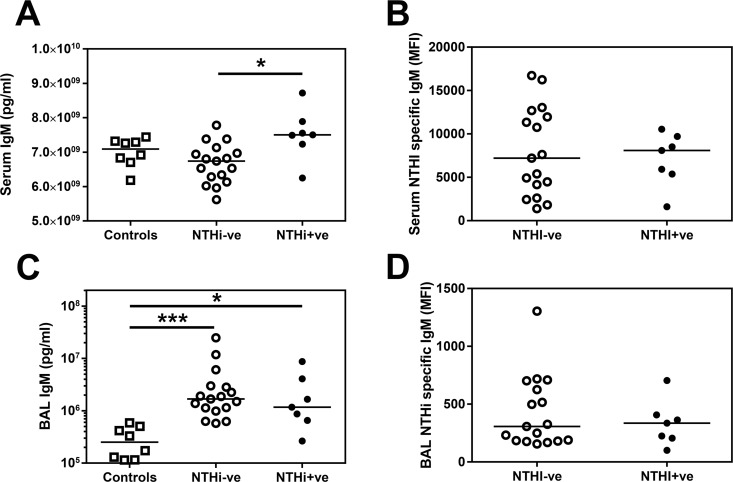
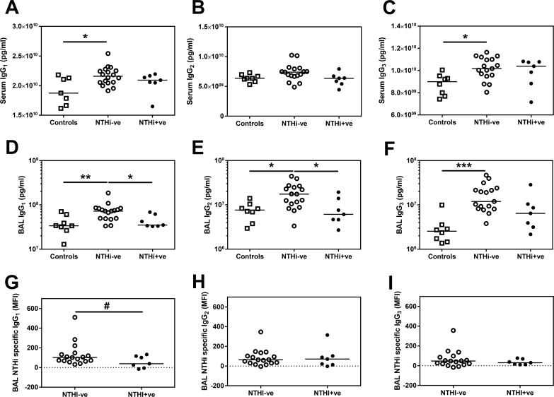
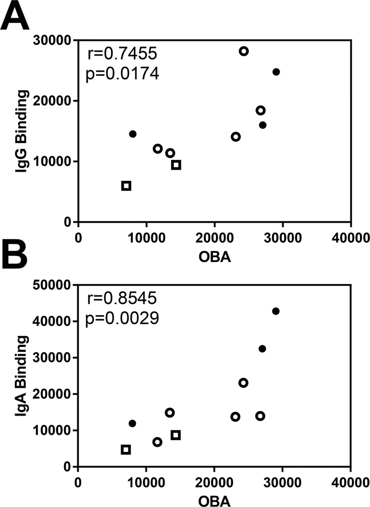
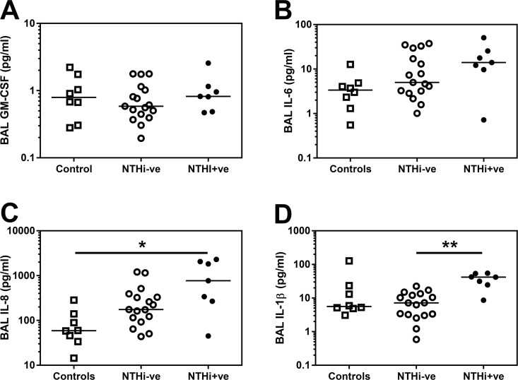
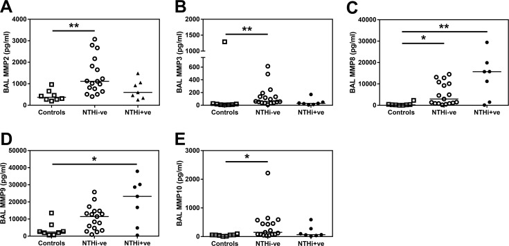
Non-typeable Haemophilus influenzae (NTHi) is a key pathogen in COPD, being associated with airway inflammation and risk of exacerbation. Why some patients are susceptible to colonisation is not understood. We hypothesised that this susceptibility may be due to a deficiency in mucosal humoral immunity. The aim of our study (NCT01701869) was to quantify the amount and specificity of antibodies against NTHi in the lungs and the associated risk of infection and inflammation in health and COPD. Phlebotomy, sputum induction and bronchoscopy were performed on 24 mild-to-moderate COPD patients and 8 age and smoking-matched controls. BAL (Bronchoalveolar lavage) total IgG1, IgG2, IgG3, IgM and IgA concentrations were significantly increased in COPD patients compared to controls. NTHi was detected in the lungs of 7 of the COPD patients (NTHi+ve-29%) and these patients had a higher median number of previous exacerbations than NTHi-ve patients as well as evidence of increased systemic inflammation. When comparing NTHi+ve versus NTHi-ve patients we observed a decrease in the amount of both total IgG1 (p = 0.0068) and NTHi-specific IgG1 (p = 0.0433) in the BAL of NTHi+ve patients, but no differences in total IgA or IgM. We observed no evidence of decreased IgG1 in the serum of NTHi+ve patients, suggesting this phenomenon is restricted to the airway. Furthermore, the NTHi+ve patients had significantly greater levels of IL-1β (p = 0.0003), in BAL than NTHi-ve COPD patients.This study indicates that the presence of NTHi is associated with reduced levels and function of IgG1 in the airway of NTHi-colonised COPD patients. This decrease in total and NTHI-specific IgG1 was associated with greater systemic and airway inflammation and a history of more frequent exacerbations and may explain the susceptibility of some COPD patients to the impacts of NTHi.


Conflict of interest statement

TGP is an employee of the GSK group of companies; he owns shares in GSK. These interests do not alter our adherence to PLOS ONE policies on sharing data and materials. The remaining authors have declared that no competing interests exist.

Figures

Fig 1
Fig 1. Volunteer recruitment CONSORT diagram.
Subjects who did not undergo bronchoscopy were excluded based on HRCT finding of other lung pathologies.
Fig 2
Fig 2. IgA and sIgA levels in BAL and serum derived from controls and COPD patients.
(A) Total IgA and (B) secretory IgA in BAL derived from two lung lobes were analysed by MSD multiplex assay and ELISA respectively and the average concentration from both lobes is presented. (C) Assessment of BAL IgA specific for the control strain of NTHi (3224A) were assessed by flow cytometry. (D) Secretory IgA in BAL derived from all volunteers are plotted against FEV1% and analysed using a Spearman’s correlation with rho and p value presented. Assessment of (E) secretory IgA in serum measured by ELISA and (F) serum IgA specific for the control strain of NTHi (3224A) were assessed by flow cytometry. Open squares indicate controls, open circles indicate NTHi-ve COPD patients, closed circles indicate NTHi+ve COPD patients. Bars represent median values and each dot represents an individual volunteer n = 8 for controls and n = 17 for NTHi-ve COPD patients and n = 7 for NTHi+ve COPD patients. Data were analysed using a Kruskal-Wallis ANOVA followed by a Dunn’s post hoc test ** p<0.01.
Fig 3
Fig 3. Total IgM but not NTHi-specific IgM are increased in the serum of NTHi+ve patients.
Total IgM in (A) serum or (C) BAL was analysed by MSD multiplex assay. (B) Serum IgM or (D) BAL IgM specific for NTHi was assessed by flow cytometry. Open circles indicate NTHi-ve COPD patients, closed circles indicate NTHi+ve patients. Bars represent median values and each dot represents an individual volunteer. n = 8 for controls, n = 17 for NTHi-ve and n = 7 for NTHi+ve patients. Data were analysed using a Kruskal-Wallis ANOVA followed by a Dunn’s post hoc test. * p<0.05, *** p<0.01
Fig 4
Fig 4. Total and NTHi-specific IgG1 are decreased in BAL from NTHi+ve patients.
Total (A) serum IgG1, (B) serum IgG2 (C) serum IgG3 was analysed by MSD multiplex assay. Total (D) IgG1, (E) IgG2 and (F) IgG3 in BAL derived from two lung lobes were analysed by MSD multiplex assay and the average concentration from both lobes is presented. Bars represent median values and each dot represents an individual volunteer n = 8 for controls, n = 17 for NTHi-ve and n = 7 for NTHi+ve patients. Data were analysed using a Kruskal-Wallis ANOVA followed by a Dunn’s post hoc test. * p<0.05, ** p<0.01, *** p<0.01. (G) BAL IgG1, (H) BAL IgG2 or (I) BAL IgG3 specific for NTHI was assessed by flow cytometry. Open squares indicate controls, open circles indicate NTHi-ve COPD patients, closed circles indicate NTHi+ve patients. Bars represent median values and each dot represents an individual volunteer n = 17 for NTHi-ve and n = 7 for NTHi+ve patients. # p<0.05 using a one tailed Mann Whitney test.
Fig 5
Fig 5. Correlation of NTHi-specific antibody binding with oxidative burst assay.
Spearman’s correlation of NTHi-specific (A) total IgG binding with IgG specific oxidative burst assay (OBA) and (B) total IgA binding with IgA specific OBA in serum with rho and p value presented. Open squares indicate controls (n = 2), open circles indicate NTHi-ve COPD patients (n = 5), closed circles indicate NTHi+ve patients (n = 3).
Fig 6
Fig 6. Inflammatory milieu is altered in NTHi+ve COPD BAL.
(A) GM-CSF (B) IL-6, (C) IL-8 and (D) IL-1β in BAL derived from two lung lobes were analysed by Luminex multiplex assay and the average concentration from both lobes is presented. Open squares indicate controls, open circles indicate NTHi-ve COPD patients, closed circles indicate NTHi+ve patients. Bars represent median values and each dot represents an individual volunteer n = 8 for controls, n = 17 for NTHi-ve and n = 7 for NTHi+ve patients. Data were analysed using a Kruskal-Wallis ANOVA followed by a Dunn’s post hoc test. * p<0.05, ** p<0.01, *** p<0.01.
Fig 7
Fig 7. MMP levels are altered in COPD BAL.
(A) MMP2, (B) MMP3, (C) MMP8, (D) MMP9 and (E) MMP10 in BAL derived from two lung lobes were analysed by Luminex multiplex assay and the average concentration from both lobes is presented. Open squares indicate controls, open circles indicate NTHi-ve COPD patients, closed circles indicate NTHi+ve patients. Bars represent median values and each dot represents an individual volunteer n = 8 for controls, n = 17 for NTHi-ve and n = 7 for NTHi+ve patients. Data were analysed using a Kruskal-Wallis ANOVA followed by a Dunn’s post hoc test. * p<0.05, ** p<0.01, *** p<0.01.

Similar articles

Cited by

References

    1. Van Eldere J, Slack MP, Ladhani S, Cripps AW. Non-typeable Haemophilus influenzae, an under-recognised pathogen. Lancet Infect Dis. 2014;14(12):1281–92. 10.1016/S1473-3099(14)70734-0 - DOI - PubMed
    1. Patel IS, Seemungal TA, Wilks M, Lloyd-Owen SJ, Donaldson GC, Wedzicha JA. Relationship between bacterial colonisation and the frequency, character, and severity of COPD exacerbations. Thorax. 2002;57(9):759–64. PubMed Central PMCID: PMCPMC1746426. 10.1136/thorax.57.9.759 - DOI - PMC - PubMed
    1. Singh R, Mackay AJ, Patel AR, Garcha DS, Kowlessar BS, Brill SE, et al. Inflammatory thresholds and the species-specific effects of colonising bacteria in stable chronic obstructive pulmonary disease. Respir Res. 2014;15:114 PubMed Central PMCID: PMCPMC4173051. 10.1186/s12931-014-0114-1 - DOI - PMC - PubMed
    1. Bafadhel M, Haldar K, Barker B, Patel H, Mistry V, Barer MR, et al. Airway bacteria measured by quantitative polymerase chain reaction and culture in patients with stable COPD: relationship with neutrophilic airway inflammation, exacerbation frequency, and lung function. Int J Chron Obstruct Pulmon Dis. 2015;10:1075–83. PubMed Central PMCID: PMCPMC4468933. 10.2147/COPD.S80091 - DOI - PMC - PubMed
    1. Sethi S, Evans N, Grant BJ, Murphy TF. New strains of bacteria and exacerbations of chronic obstructive pulmonary disease. N Engl J Med. 2002;347(7):465–71. Epub 2002/08/16. 10.1056/NEJMoa012561 - DOI - PubMed

MeSH terms