

Environmental Free-Living Amoebae Isolated from Soil in Khon Kaen, Thailand, Antagonize Burkholderia pseudomallei
- PMID: 27898739
- PMCID: PMC5127566
- DOI: 10.1371/journal.pone.0167355
Environmental Free-Living Amoebae Isolated from Soil in Khon Kaen, Thailand, Antagonize Burkholderia pseudomallei
Abstract
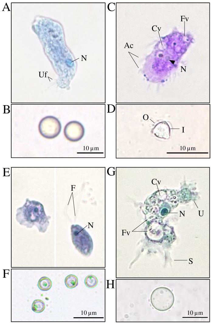
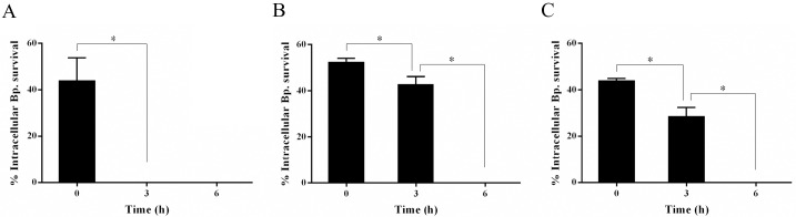
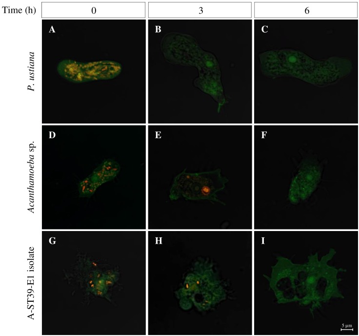
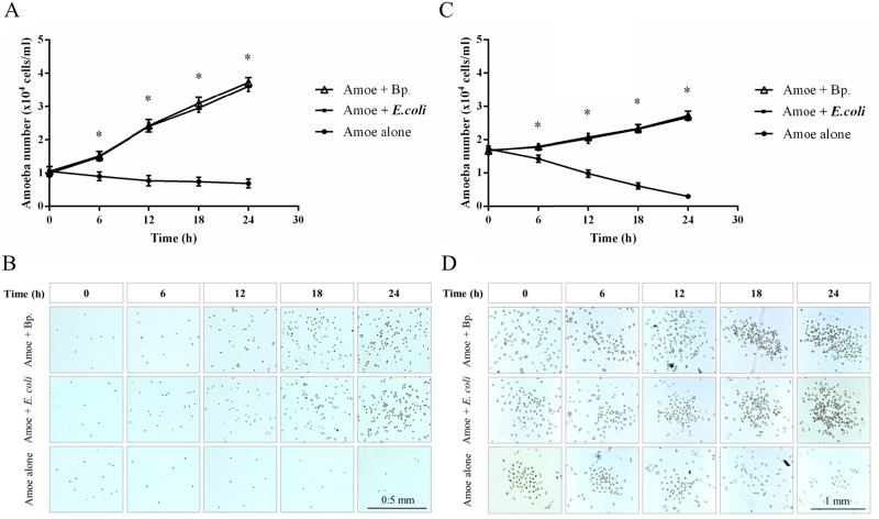
Presence of Burkholderia pseudomallei in soil and water is correlated with endemicity of melioidosis in Southeast Asia and northern Australia. Several biological and physico-chemical factors have been shown to influence persistence of B. pseudomallei in the environment of endemic areas. This study was the first to evaluate the interaction of B. pseudomallei with soil amoebae isolated from B. pseudomallei-positive soil site in Khon Kaen, Thailand. Four species of amoebae, Paravahlkampfia ustiana, Acanthamoeba sp., Naegleria pagei, and isolate A-ST39-E1, were isolated, cultured and identified based on morphology, movement and 18S rRNA gene sequence. Co-cultivation combined with a kanamycin-protection assay of B. pseudomallei with these amoebae at MOI 20 at 30°C were evaluated during 0-6 h using the plate count technique on Ashdown's agar. The fate of intracellular B. pseudomallei in these amoebae was also monitored by confocal laser scanning microscopy (CLSM) observation of the CellTracker™ Orange-B. pseudomallei stained cells. The results demonstrated the ability of P. ustiana, Acanthamoeba sp. and isolate A-ST39-E1 to graze B. pseudomallei. However, the number of internalized B. pseudomallei substantially decreased and the bacterial cells disappeared during the observation period, suggesting they had been digested. We found that B. pseudomallei promoted the growth of Acanthamoeba sp. and isolate A-ST39-E1 in co-cultures at MOI 100 at 30°C, 24 h. These findings indicated that P. ustiana, Acanthamoeba sp. and isolate A-ST39-E1 may prey upon B. pseudomallei rather than representing potential environmental reservoirs in which the bacteria can persist.
Conflict of interest statement
The authors have declared that no competing interests exist.
Figures
Similar articles
-
Soil physicochemical properties related to the presence of Burkholderia pseudomallei.Trans R Soc Trop Med Hyg. 2008 Dec;102 Suppl 1:S5-9. doi: 10.1016/S0035-9203(08)70003-8. Trans R Soc Trop Med Hyg. 2008. PMID: 19121688
-
Epidemiology of Burkholderia pseudomallei in Thailand.Am J Trop Med Hyg. 1999 Mar;60(3):458-61. doi: 10.4269/ajtmh.1999.60.458. Am J Trop Med Hyg. 1999. PMID: 10466977
-
Antibacterial activity of chitosan against Burkholderia pseudomallei.Microbiologyopen. 2018 Feb;7(1):e00534. doi: 10.1002/mbo3.534. Epub 2017 Nov 27. Microbiologyopen. 2018. PMID: 29178614 Free PMC article.
-
Burkholderia pseudomallei: the unbeatable foe?Southeast Asian J Trop Med Public Health. 1998 Jun;29(2):410-5. Southeast Asian J Trop Med Public Health. 1998. PMID: 9886137 Review.
-
Vannella primoblina n. sp. - an unusual species of the genus Vannella (Amoebozoa, Discosea, Vannellida) with pronounced dorsal ridges and folds.Eur J Protistol. 2021 Feb;77:125757. doi: 10.1016/j.ejop.2020.125757. Epub 2020 Nov 20. Eur J Protistol. 2021. PMID: 33307358 Review.
Cited by
-
Human Melioidosis.Clin Microbiol Rev. 2020 Mar 11;33(2):e00006-19. doi: 10.1128/CMR.00006-19. Print 2020 Mar 18. Clin Microbiol Rev. 2020. PMID: 32161067 Free PMC article. Review.
-
An Evolutionary Arms Race Between Burkholderia pseudomallei and Host Immune System: What Do We Know?Front Microbiol. 2021 Jan 21;11:612568. doi: 10.3389/fmicb.2020.612568. eCollection 2020. Front Microbiol. 2021. PMID: 33552023 Free PMC article. Review.
-
Burkholderia pseudomallei Adaptation for Survival in Stressful Conditions.Biomed Res Int. 2018 May 27;2018:3039106. doi: 10.1155/2018/3039106. eCollection 2018. Biomed Res Int. 2018. PMID: 29992136 Free PMC article. Review.
-
Genetic variation associated with infection and the environment in the accidental pathogen Burkholderia pseudomallei.Commun Biol. 2019 Nov 22;2:428. doi: 10.1038/s42003-019-0678-x. eCollection 2019. Commun Biol. 2019. PMID: 31799430 Free PMC article.
-
Burkholderia pseudomallei biofilm resists Acanthamoeba sp. grazing and produces 8-O-4'-diferulic acid, a superoxide scavenging metabolite after passage through the amoeba.Sci Rep. 2023 Oct 3;13(1):16578. doi: 10.1038/s41598-023-43824-1. Sci Rep. 2023. PMID: 37789212 Free PMC article.
References
MeSH terms
Substances
LinkOut - more resources
Full Text Sources
Other Literature Sources
