A functional magnetic resonance imaging investigation of visual hallucinations in the human striate cortex
- PMID: 27899123
- PMCID: PMC5126868
- DOI: 10.1186/s12993-016-0115-y
A functional magnetic resonance imaging investigation of visual hallucinations in the human striate cortex
Abstract
Purpose: Human beings frequently experience fear, phobia, migraine and hallucinations, however, the cerebral mechanisms underpinning these conditions remain poorly understood. Towards this goal, in this work, we aim to correlate the human ocular perceptions with visual hallucinations, and map them to their cerebral origins.
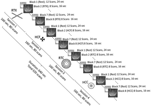
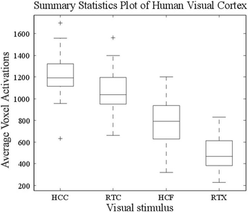
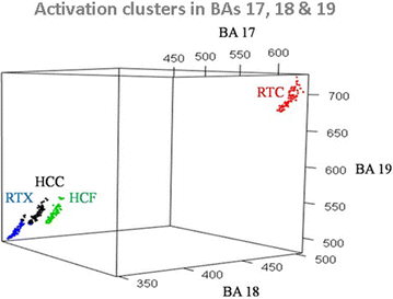
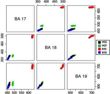
Methods: An fMRI study was performed to examine the visual cortical areas including the striate, parastriate and peristriate cortex in the occipital lobe of the human brain. 24 healthy subjects were enrolled and four visual patterns including hallucination circle (HCC), hallucination fan (HCF), retinotopy circle (RTC) and retinotopy cross (RTX) were used towards registering their impact in the aforementioned visual related areas. One-way analysis of variance was used to evaluate the significance of difference between induced activations. Multinomial regression and and K-means were used to cluster activation patterns in visual areas of the brain.
Results: Significant activations were observed in the visual cortex as a result of stimulus presentation. The responses induced by visual stimuli were resolved to Brodmann areas 17, 18 and 19. Activation data clustered into independent and mutually exclusive clusters with HCC registering higher activations as compared to HCF, RTC and RTX.
Conclusions: We conclude that small circular objects, in rotation, tend to leave greater hallucinating impressions in the visual region. The similarity between observed activation patterns and those reported in conditions such as epilepsy and visual hallucinations can help elucidate the cortical mechanisms underlying these conditions. Trial Registration 1121_GWJUNG.
Keywords: Brodmann area; Functional magnetic resonance imaging (fMRI); K-means clustering; Logistic regression; Visual cortex; Visual hallucinations.
Figures


References
-
- Finger S. Origins of neuroscience: a history of explorations into brain function. New York: Oxford University Press; 1994.
-
- Garey LJ. Brodmann’s localisation in the cerebral cortex—the principles of comparative localisation in the cerebral cortex based on cytoarchitectonics. New York: Springer Science; 2006.
-
- Ahmad F, et al. A shrinkage method for causal network detection of brain regions. Imag Sys Technol. 2013;23(2):140–146. doi: 10.1002/ima.22047. - DOI
MeSH terms
LinkOut - more resources
Full Text Sources
Other Literature Sources
Medical

