. 2017 Jan:85:399-411.
doi: 10.1016/j.biopha.2016.11.043. Epub 2016 Nov 26.

Estrogen and thyroid cancer is a stem affair: A preliminary study

Affiliations

Estrogen and thyroid cancer is a stem affair: A preliminary study

Mariangela Zane et al. Biomed Pharmacother. 2017 Jan.

Abstract

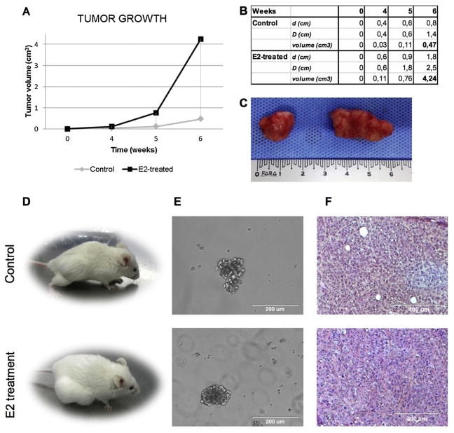
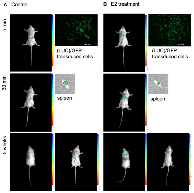
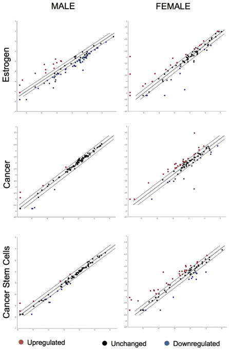
Gender influences Papillary Thyroid Cancer (PTC) with an incidence of 3:1 when comparing women to men with different aggressiveness. This gender discrepancy suggests some role of sex hormones in favoring the malignant progression of thyroid tissue to cancer. Estrogens are known to promote Stem Cell self-renewal and, therefore, may be involved in tumor initiation. The goals of these studies are to investigate the underlying causes of gender differences in PTC by studying the specific role of estrogens on tumor cells and their involvement within the Cancer Stem Cell (CSC) compartment. Exposure to 1nmoll-1 Estradiol for 24h promotes growth and maintenance of PTC Stem Cells, while inducing dose-dependent cellular proliferation and differentiation following Estradiol administration. Whereas mimicking a condition of hormonal imbalance led to an opposite phenotype compared to a continuous treatment. In vivo we find that Estradiol promotes motility and tumorigenicity of CSCs. Estradiol-treated mice inoculated with Thyroid Cancer Stem Cell-enriched cells developed larger tumor masses than control mice. Furthermore, Estradiol-pretreated Cancer Stem cells migrated to distant organs, while untreated cells remained circumscribed. We also find that the biological response elicited by estrogens on Papillary Thyroid Cancer in women differed from men in pathways mediated. This could explain the gender imbalance in tumor incidence and development and could be useful to develop gender specific treatment of (PTC).

Keywords: Cancer signaling; Cancer stem cells; Estrogen; Gender medicine; Thyroid cancer.


Conflict of interest statement

Declaration of interest The authors declare that there is no conflict of interest that could be perceived as prejudicing the impartiality of the research reported.

Figures

Fig. 1
Fig. 1
Thyroid tumor bulk contains stem-like cells. (A) Percentage of ALDHhigh cells within K1 and BCPAP cell lines: 23.6% in K1, 4.87% in BCPAP. (B) Immunofluorescence anatlysis of SOX2, OCT3, BETA-CATENIN, and TSHR on primary cell cultures (zoom 40×; confocal microscope). (C) Optimization of isolation methods. Cell culture maintained in attachment and ultra-low adhesion, in the presence or absence of FBS (magnitude 10×; optical microscope).
Fig. 2
Fig. 2
BMP pathway inhibition acts with FGF2 in maintenance of stemness in vitro. (A) Histological examination of paraffined PTC samples (magnitude 40×, 63×, 40×, respectively; optical microscope). (B) CSC-enriched cell cultures isolated from the same PTC samples, able to maintain the double (adherent and floating) population in culture (magnitude 20×; optical microscope). (C) FACS analysis on CSC-enriched cell cultures for SOX2, OCT3, PAX8, TTF1, ERA, and ERB after treatment with FGF2 and NOGGIN. Conditions: control; 1 ug/100 mL FGF2; 10 ug/100 mL NOGGIN + 1 ug/100 mL FGF2; 10 ug/100 mL NOGGIN + 4 ug/100 mL FGF2. 48 h treatment.
Fig. 3
Fig. 3
Estrogen treatment enhances stem markers expression in a time- and dose-dependent manner. (A) Immunofluorescence analysis on BCPAP for SOX2, OCT3, NANOG, PAX8, TTF1, TSHR, TG, TPO, NIS, BETA-CATENIN, ERA, and ERB after E2 treatment at 1 and 10 nmol l−1 for 24 h and 48 h (magnitude 40×, confocal microscope). (B) Same immunofluorescence analysis on CSC-enriched patient cell cultures. (C) FACS analysis on CSC-enriched cell cultures for SOX2, OCT3, PAX8, TTF1, ERA, and ERB after E2 treatment.
Fig. 4
Fig. 4
A continuous presence of Estradiol has an opposite effect compared to a hormonal imbalance simulation. (A) Morphological evaluation of CSC-enriched cell cultures after 1 and 10 nmol l−1 continuous E2 treatment, and with hormonal imbalance simulation (magnitude 10×; optical microscope). (B) Mean Fluoresce Index of FACS analysis for SOX2, OCT3, NANOG, ERA, ERB, and GPER1. UNTREAT = control; E2 TREAT = cells after 7-days treatment; STOPPING TREAT = cells after stopping treatment for further 7 days. * = P < 0.005.
Fig. 5
Fig. 5
Host estrogen therapy enhances the tumorigenic potential of thyroid CSCs. (A) Diagram of tumor volume growth with or without host E2 therapy. 4,24 vs. 0.47 cm3 mean, * = P < 0.05. (B) Macroscopic analysis of xenograft tumors. Untreated vs E2-treated. (C) Subcutaneous xenografts obtained by the injection of CSC-primary culture cells (1.5 × 10⁶) in immunocompromised NOD/SCID Il2rg -/- female mice, alone or in combination with estrogen therapy. Untreated vs E2-treated. (D) Thyroidspheres in culture after xenograft tumor digestion (magnitude 20×, optical microscope). Untreated vs E2-treated. (E) H&E staining of paraffined xenograft tumors (magnitude 10×, optical microscope). Untreated vs E2-treated.
Fig. 6
Fig. 6
E2-treated CSCs empower their migratory activity in vivo. Intrasplenic injection in NOD/SCID female mice of 3 ×10⁵ luciferase (LUC)/GFP-transduced cells, untreated or treated with 1 nmol l−1 E2 for 24 h (magnitude 10×, EVOS™, Thermo Fischer, Waltham, Massachussetts). Analysis of migratory capacity at time: 0 min; 30 min; 5 weeks. Biospace.
Fig. 7
Fig. 7
Estrogens perform their function in a different way among women and men. Estrogen, cancer, and cancer stem cell pathway analysis for female and male CSC-enriched cell cultures. The scatter plot compares the normalized expression of 252 genes on the array between two groups (untreated and E2-treated).

References

    1. Ahmed RA, Aboelnaga EM. Thyroid cancer in Egypt: histopathological criteria, correlation with survival and oestrogen receptor protein expression. Pathol Oncol Res. 2015;21:793–802. - PubMed
    1. Antico-Arciuch VG, Dima M, Liao XH, Refetoff S, Di Cristofano A. Cross-talk between PI3K and estrogen in the mouse thyroid predisposes to the development of follicular carcinomas with a higher incidence in females. Oncogene. 2010;29:5678–5686. - PMC - PubMed
    1. Antico Arciuch VG, Di Cristofano A. Estrogen signaling and thyrocyte proliferation. In: Ward DL, editor. Thyroid and Parathyroid Diseases-New insights into Some Old and Some New Issues. 2012.
    1. Braganza MZ, de Gonzalez AB, Schonfeld SJ, Wentzensen N, Brenner AV, Kitahara CM. Benign breast and gynecologic conditions, reproductive and hormonal factors, and risk of thyroid cancer. Cancer Prev Res (Phila) 2014;7:418–425. - PMC - PubMed
    1. Caini S, Gibelli B, Palli D, Saieva C, Ruscica M, Gandini S. Menstrual and reproductive history and use of exogenous sex hormones and risk of thyroid cancer among women: a meta-analysis of prospective studies. Cancer Causes Control. 2015;26:511–518. - PubMed