. 2017 Mar 1:114:123-134.
doi: 10.1016/j.neuropharm.2016.11.021. Epub 2016 Nov 27.

Sex differences in responses of the basolateral-central amygdala circuit to alcohol, corticosterone and their interaction

Affiliations

Sex differences in responses of the basolateral-central amygdala circuit to alcohol, corticosterone and their interaction

Marian L Logrip et al. Neuropharmacology. .

Abstract

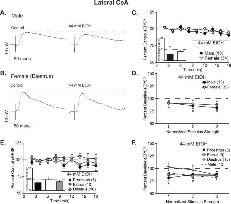
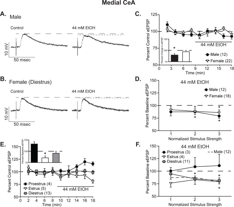
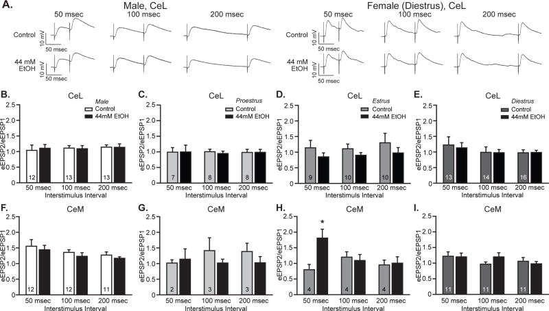
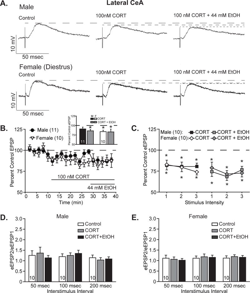
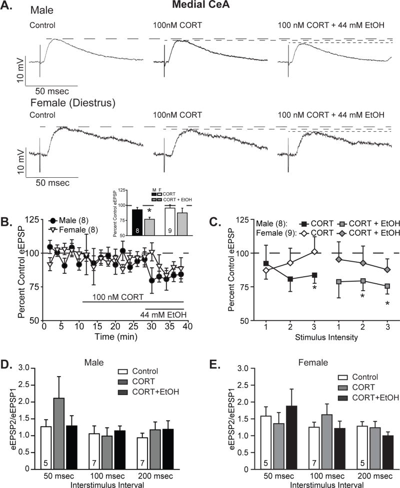
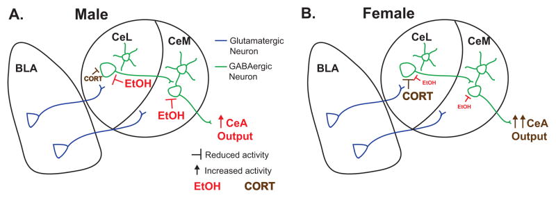
Alcohol use disorders are chronically relapsing conditions that pose significant health challenges for our society. Stress is a prevalent trigger of relapse, particularly for women, yet the mechanisms by which alcohol and stress interact, and how this differs between males and females, remain poorly understood. The glutamatergic circuit connecting the basolateral (BLA) and central (CeA) nuclei of the amygdala is a likely locus for such adaptations, yet the impact of alcohol, corticosterone and their interaction on this circuit has been understudied. In particular, no studies have addressed sex differences in these effects or potential differential responses between the lateral and medial subdivisions of the central nucleus. Thus, we assessed the effects of alcohol and corticosterone treatments on BLA-evoked compound glutamatergic responses in medial and lateral CeA neurons from male and female rats. We observed minimal differences between medial and lateral CeA responses to alcohol and corticosterone in male rats, which were primarily sensitive to alcohol-induced inhibition of glutamatergic postsynaptic potentials. Unlike male neurons, cells from female rats displayed reduced sensitivity to alcohol's inhibitory effects. In addition, female neurons diverged in their sensitivity to corticosterone, with lateral CeA neuronal responses significantly blunted following corticosterone treatment and medial CeA neurons largely unchanged by corticosterone or subsequent co-application of alcohol. Together these data highlight striking differences in how male and female amygdala respond to alcohol and the stress hormone corticosterone, factors which may impact differential susceptibility of the sexes to alcohol- and stress-related disorders.

Keywords: Amygdala; Electrophysiology; Ethanol; Sex differences; Stress.


Figures

Figure 1
Figure 1. Ethanol acutely reduced the amplitude of BLA-evoked compound glutamatergic EPSPs in the lateral CeA
A,B. Representative evoked glutamatergic-EPSPs (eEPSPs) in CeL, at baseline (Control) and during 44 mM ethanol (EtOH) superfusion onto slices obtained from male (A) and diestrus female (B) rats. C. Time course of treatment effects, with ethanol application following an 8-min baseline. Inset shows quantification of the peak alcohol-induced change in eEPSPs over a 4-minute bin beginning not less than 6 min into ethanol application in males vs. all females. Histograms depict mean ± standard error percent change in eEPSPs relative to control. D. Quantification of ethanol’s effect on I/O responses to the 3 middle intensity stimuli in males vs. all females. Data are depicted as mean ± standard error percent change in eEPSP after ethanol treatment, normalized to control. E. Time course of treatment effects by estrous cycle, with ethanol application following an 8-min baseline. No significant effects were observed in females based on estrous cycle status at the time of euthanasia. Histograms (inset) depict mean ± standard error percent changes in peak alcohol effect relative to control. F. Estrous cycle impacts stimulus responsiveness as measured by the I/O relationship at 3 stimuli of increasing intensity. Data are depicted as mean ± standard error percent change in eEPSP relative to control. n’s = 8–34, as listed on each panel of the graph; cells becoming unstable after drug wash-on were excluded from I/O analyses. *p<0.05 relative to control (one-sample t-test).
Figure 2
Figure 2. Ethanol acutely reduced the amplitude of BLA-evoked compound glutamatergic EPSPs in the medial CeA, particularly in males
A,B. Representative evoked glutamatergic-EPSPs (eEPSPs) in CeM, at baseline (Control) and during 44 mM ethanol (EtOH) superfusion onto slices obtained from male (A) and diestrus female (B) rats. C. Time course of treatment effects, with ethanol application following an 8-min baseline. Inset shows quantification of the peak ethanol-induced change in eEPSP over a 4-minute bin beginning not less than 6 min into ethanol application in males vs. all females. Data are depicted as mean ± standard error percent change in eEPSP relative to control. D. Quantification of ethanol’s effect on I/O responses to 3 intermediate intensity stimuli in males vs. all females. Data are depicted as mean ± standard error percent change in eEPSP after ethanol application, normalized to control. E. Time course of treatment effects by estrous cycle, with ethanol superfusion ollowing an 8-min baseline. Estrous cycle affects ethanol’s modulation of peak eEPSP amplitude. Histograms depict mean ± standard error percent changes in peak ethanol effect relative to control. F. Estrous cycle impacts CeM neurons’ stimulus responsiveness, as measured by the I/O relationship at 3 intermediate intensity stimuli. Data are depicted as mean ± standard error percent change in eEPSP relative to control. n’s = 3–22, as listed on each panel of the graph; cells becoming unstable after drug wash-on were excluded from I/O analyses. *p<0.05 relative to control (one-sample t-test).
Figure 3
Figure 3. Ethanol did not significantly alter paired pulse ratios in lateral or medial CeA neurons of either sex
A. Representative traces of paired pulse EPSPs evoked by two pulses delivered 50 msec (left), 100 msec (center) or 200 msec (right) apart, assessed at baseline (Control) and during superfusion of 44 mM ethanol (EtOH) onto slices from male and female rats. B-I. Quantification of the relationship between peak responses to the paired stimuli (paired pulse ratio), calculated as the ratio between the second stimulus response (eEPSP2) and the first stimulus responses (eEPSP1). Histograms depict mean ± standard error paired pulse ratios for CeL male (B) and proestrus (C), estrus (D), and diestrus females (E) and CeM male (F) and proestrus (G) estrus (H), and diestrus females (I) cells. n’s = 10–16, as listed on the graph panels.
Figure 4
Figure 4. Corticosterone acutely reduced lateral CeA EPSPs and occluded further ethanol effects in females, while males responded more to ethanol
A. Representative evoked glutamatergic-EPSPs (eEPSPs) in CeL, at baseline (Control) and during superfusion of 100 nM corticosterone (CORT) and subsequent co-application of 44 mM ethanol (EtOH) onto slices obtained from male (top) and diestrus female (bottom) rats. B. Time course of treatment effects, with CORT application following an 8-min baseline and alcohol co-application beginning 20 min after CORT application. Inset shows quantification of the peak CORT- and ethanol-induced changes in eEPSPs over a 4-minute bin beginning not less than 15 min into CORT treatment and not less than 6 min into ethanol co-application. Histograms depict mean ± standard error percent change in eEPSP relative to control. C. Quantification of CORT and ethanol effects on I/O responses to 3 intermediate intensity stimuli in males (squares) vs. diestrus females (diamonds). Data are depicted as mean ± standard error percent change in eEPSP after CORT treatment (black and white shapes) and subsequent ethanol co-application (gray shapes), normalized to control. D–E. Histograms depict mean ± standard error paired pulse ratios for male (D) and female (E) cells. n’s = 9–11, as listed on the graph panels; cells becoming unstable after drug wash-on were excluded from I/O analyses. *p<0.05 relative to control (one-sample t-test).
Figure 5
Figure 5. Corticosterone minimally affects medial CeA responses but blocks ethanol’s effects in females, but not males
A. Representative evoked glutamatergic-EPSPs (eEPSPs) in CeM at baseline (Control) and during superfusion of 100 nM corticosterone (CORT) and subsequent co-application of 44 mM ethanol (EtOH) onto slices obtained from male (top) and diestrus female (bottom) rats. B. Time course of treatment effects, with CORT treatment following an 8-min baseline and alcohol co-application beginning 20 min after CORT application. Inset shows quantification of the peak CORT and ethanol-induced changes in eEPSPs over a 4-minute bin beginning not less than 15 min into CORT treatment and not less than 6 min into ethanol co-application. Histograms depict mean ± standard error percent change in eEPSP relative to control. C. Quantification of CORT and ethanol effects on I/O responses to 3 intermediate intensity stimuli in males (squares) vs. diestrus females (diamonds). Data are depicted as mean ± standard error percent change in eEPSP after CORT treatment (black and white shapes) and subsequent ethanol co-application (gray shapes), normalized to control. D–E. Histograms depict mean ± standard error paired pulse ratios for male (D) and female (E) cells. n’s = 5–9, as listed on the graph panels; cells becoming unstable after drug wash-on were excluded from I/O analyses, and one female cell with incomplete data was omitted from the time course. *p<0.05 relative to control (one-sample t-test).
Figure 6
Figure 6. Schematic representation of observed sex differences
Ethanol (EtOH) and corticosterone (CORT) differentially modulate male (A) and female (B) lateral (CeL) and medial (CeM) central amygdala synaptic responses to basolateral amygdala (BLA) stimulation. (A) In males, EtOH reduces EPSPs, whether the sole drug treatment or following CORT treatment, in both CeL and CeM, while CORT has a reduced effect on eEPSPs relative to EtOH and to CORT’s effects in females. (B) In females, CORT significantly reduces CeL, but not CeM, eEPSPs, whereas EtOH has a reduced ability to inhibit eEPSPs in females vs. males that is blocked in the presence of CORT.

References

    1. Aggleton JP, editor. The Amygdala: A Functional Analysis. Oxford University Press; 2000.
    1. Almeida OF, Shoaib M, Deicke J, Fischer D, Darwish MH, Patchev VK. Gender differences in ethanol preference and ingestion in rats. The role of the gonadal steroid environment. J Clin Invest. 1998;101:2677–2685. - PMC - PubMed
    1. Atkinson HC, Waddell BJ. Circadian variation in basal plasma corticosterone and adrenocorticotropin in the rat: sexual dimorphism and changes across the estrous cycle. Endocrinology. 1997;138:3842–3848. - PubMed
    1. Cassell MD, Freedman LJ, Shi C. The intrinsic organization of the central extended amygdala. Ann N Y Acad Sci. 1999;877:217–241. - PubMed
    1. Cassell MD, Gray TS. Morphology of peptide-immunoreactive neurons in the rat central nucleus of the amygdala. J Comp Neurol. 1989;281:320–333. - PubMed

Publication types