. 2016 Dec 12;213(13):3075-3086.
doi: 10.1084/jem.20160888. Epub 2016 Nov 29.

Antigen-dependent competition shapes the local repertoire of tissue-resident memory CD8+ T cells

Affiliations

Antigen-dependent competition shapes the local repertoire of tissue-resident memory CD8+ T cells

Andreas Muschaweckh et al. J Exp Med. .

Abstract

Tissue-resident memory CD8+ T cells (TRM) constitute a major component of the immune-surveillance system in nonlymphoid organs. Local, noncognate factors are both necessary and sufficient to support the programming of TRM cell fate in tissue-infiltrating T cells. Recent evidence suggests that TCR signals received in infected nonlymphoid tissues additionally contribute to TRM cell formation. Here, we asked how antigen-dependent pathways influence the generation of skin-resident memory T cells that arise from a polyclonal repertoire of cells induced by infection with an antigenically complex virus and recombinant vaccine vector. We found that CD8+ T cells of different specificities underwent antigen-dependent competition in the infected tissue, which shaped the composition of the local pool of TRM cells. This local cross-competition was active for T cells recognizing antigens that are coexpressed by infected cells. In contrast, TRM cell development remained largely undisturbed by the presence of potential competitors when antigens expressed in the same tissue were segregated through infection with antigenically distinct viral quasispecies. Functionally, local cross-competition might serve as a gatekeeping mechanism to regulate access to the resident memory niche and to fine-tune the local repertoire of antiviral TRM cells.


Figures

Figure 1.
Figure 1.
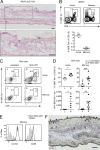
Localized MVA skin infection induces bona fide TRM cells. (A) Localization of virus-infected cells in ear skin 16 h after intraepidermal infection with MVA-LacZ (107 IU), as visualized by X-Gal staining. Bar, 100 µm. Shown is a representative staining of at least three independent experiments. (B) Representative plots (top) and percentage (bottom) of B8R-specific CD8+ T cells, in the spleen 7 d (Acute) or >70 d (Memory) pi of ear skin with MVA-WT, as determined by tetramer staining. (C) Representative plots showing the B8R-specific CD8+ T cell fraction among CD45+ cells in untreated left (L) or MVA-WT–infected right (R) ears during the acute or memory phase. (D) Frequency (top) and absolute number (bottom) of B8R-specific CD8+ T cells in the indicated ears during the acute and memory phase. (E) CD103 and CD69 expression on B8R-specific memory CD8+ T cells isolated >70 d pi from previously MVA-WT–infected ears (black histogram) or the spleen (gray shaded histogram). (F) Distribution of adoptively transferred OVA-specific CD45.1+ OT-I cells (arrows) in ear skin 50 d after local infection with MVA-OVA, as detected by immunohistochemical staining of the CD45.1 (arrowheads). Bar, 100 µm. Shown is a representative staining of at least three independent experiments. Data are derived from one experiment (Acute, n = 5; representative of two independent experiments) or were pooled from three independent experiments (Memory, day 70–90, n = 8). Bars show mean + SEM. *, P < 0.05; **, P < 0.01; ***, P < 0.001 (paired Student’s t test).
Figure 2.
Figure 2.
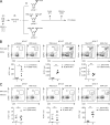
Local cognate antigen expression dramatically amplifies generation of antiviral skin TRM cells. (A) Mice were adoptively transferred with 5 × 10⁴ OVA-specific CD45.1+ OT-I cells 1 d before infection (i.v.) with MVA-OVA. Immediately after i.v. infection, mice were divided into three groups. Group 1 was mock infected (PBS mock) on the right ear (R). Group 2 was mock infected (PBS mock) on the left ear (L) and infected with MVA-WT on the right ear (R). Group 3 was infected on the left (L) and right ear (R) with MVA-WT and MVA-OVA, respectively. 7 d (Acute) and 40 d (Memory) pi, total numbers of OT-I cells were determined in differentially inoculated ears. (B and C) Representative plots depicting the frequency of OT-I cells (among CD45+ cells) in the indicated skin region 7 d (B) or 40 d (C) pi (top). Total number of OT-I cells in each ear 7 d (B) and 40 d (C) after inoculation (bottom). Data are derived from one experiment representative of at least two independent experiments (Acute, n = 4; Memory, n = 5). Dots in graphs show individual mice and bars show mean + SEM. *, P < 0.05; **, P < 0.01 (paired Student’s t test).
Figure 3.
Figure 3.
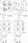
Local antigen-dependent competition shapes the pool of skin-resident memory T cells. (A) OT-I–recipient mice were primed i.v. with MVA-OVA and immediately thereafter infected on the left (L) or right (R) ear pinna with MVA-WT or MVA-OVA, respectively (Fig. 2 A, group 3). Percentage (representative plots and top graphs, right) and absolute number (bottom graphs, right) of CD45.1+ OT-I cells and endogenous B8R-specific T cells among CD8+ T cells in the differentially infected skin 7 d (Acute) and 40 d (Memory) pi. Data were pooled from two to three independent experiments (Acute, n = 9; Memory, n = 7). (B) Percentage (representative blots and top graph) and absolute number (bottom graph) of endogenous OVA- and B8R-specific T cells among CD8+ T cells in MVA-WT and MVA-OVA–infected ears 40 d pi of mice as in A, but without prior transfer of OT-I. Data were pooled from two independent experiments (n = 10). (C) Fraction (representative blots and top graph) and absolute number (bottom graph) of OVA- and B8R-specific T cells among CD8+ T cells in MVA-OVA ΔB8R–infected versus MVA-OVA–infected skin 40 d pi. Data were pooled from two independent experiments (n = 12). Dots in graphs depict individual mice and bars show mean + SEM. *, P < 0.05; **, P < 0.01; ***, P < 0.001; ****, P < 0.001 (paired Student’s t test).
Figure 4.
Figure 4.
Local cross-competition in response to coexpressed viral antigens. (A) OT-I–recipient mice were primed i.v. with MVA-OVA and co-infected on the left ear (L) with a 1:1 mixture of mutant MVA-OVA ΔB8R (expressing OVA but not B8) and MVA-WT (lacking OVA but expressing B8) for separate delivery of OVA and B8 into the skin. For antigen (Ag) co-delivery, contralateral (right) ears (R) were co-infected with a 1:1 mix of MVA-OVA (expressing both OVA and B8) and MVA ΔB8R (lacking both OVA and B8). (B) Percentages of OT-I and endogenous B8R-specific CD8+ T cells in the indicated skin regions 7 d (Acute) and 40 d (Memory) pi. Data are derived from one experiment (Acute, n = 4, representative of two independent experiments) or were pooled from two independent experiments (Memory, n = 7). Dots in graphs show individual mice and horizontal bars show mean + SEM. **, P < 0.01; ***, P < 0.001 (paired Student’s t test).

References

    1. Ariotti S., Beltman J.B., Chodaczek G., Hoekstra M.E., van Beek A.E., Gomez-Eerland R., Ritsma L., van Rheenen J., Marée A.F., Zal T., et al. . 2012. Tissue-resident memory CD8+ T cells continuously patrol skin epithelia to quickly recognize local antigen. Proc. Natl. Acad. Sci. USA. 109:19739–19744. 10.1073/pnas.1208927109 - DOI - PMC - PubMed
    1. Bins A.D., Jorritsma A., Wolkers M.C., Hung C.F., Wu T.C., Schumacher T.N., and Haanen J.B.. 2005. A rapid and potent DNA vaccination strategy defined by in vivo monitoring of antigen expression. Nat. Med. 11:899–904. 10.1038/nm1264 - DOI - PubMed
    1. Bromley S.K., Thomas S.Y., and Luster A.D.. 2005. Chemokine receptor CCR7 guides T cell exit from peripheral tissues and entry into afferent lymphatics. Nat. Immunol. 6:895–901. 10.1038/ni1240 - DOI - PubMed
    1. Casey K.A., Fraser K.A., Schenkel J.M., Moran A., Abt M.C., Beura L.K., Lucas P.J., Artis D., Wherry E.J., Hogquist K., et al. . 2012. Antigen-independent differentiation and maintenance of effector-like resident memory T cells in tissues. J. Immunol. 188:4866–4875. 10.4049/jimmunol.1200402 - DOI - PMC - PubMed
    1. Debes G.F., Arnold C.N., Young A.J., Krautwald S., Lipp M., Hay J.B., and Butcher E.C.. 2005. Chemokine receptor CCR7 required for T lymphocyte exit from peripheral tissues. Nat. Immunol. 6:889–894. 10.1038/ni1238 - DOI - PMC - PubMed

Publication types