. 2017 Jan 9;45(1):1-14.
doi: 10.1093/nar/gkw1046. Epub 2016 Nov 28.

Division of labor among Mycobacterium smegmatis RNase H enzymes: RNase H1 activity of RnhA or RnhC is essential for growth whereas RnhB and RnhA guard against killing by hydrogen peroxide in stationary phase

Affiliations

Division of labor among Mycobacterium smegmatis RNase H enzymes: RNase H1 activity of RnhA or RnhC is essential for growth whereas RnhB and RnhA guard against killing by hydrogen peroxide in stationary phase

Richa Gupta et al. Nucleic Acids Res. .

Abstract

RNase H enzymes sense the presence of ribonucleotides in the genome and initiate their removal by incising the ribonucleotide-containing strand of an RNA:DNA hybrid. Mycobacterium smegmatis encodes four RNase H enzymes: RnhA, RnhB, RnhC and RnhD. Here, we interrogate the biochemical activity and nucleic acid substrate specificity of RnhA. We report that RnhA (like RnhC characterized previously) is an RNase H1-type magnesium-dependent endonuclease with stringent specificity for RNA:DNA hybrid duplexes. Whereas RnhA does not incise an embedded mono-ribonucleotide, it can efficiently cleave within tracts of four or more ribonucleotides in duplex DNA. We gained genetic insights to the division of labor among mycobacterial RNases H by deleting the rnhA, rnhB, rnhC and rnhD genes, individually and in various combinations. The salient conclusions are that: (i) RNase H1 activity is essential for mycobacterial growth and can be provided by either RnhC or RnhA; (ii) the RNase H2 enzymes RnhB and RnhD are dispensable for growth and (iii) RnhB and RnhA collaborate to protect M. smegmatis against oxidative damage in stationary phase. Our findings highlight RnhC, the sole RNase H1 in pathogenic mycobacteria, as a candidate drug discovery target for tuberculosis and leprosy.


Figures

Figure 1.
Figure 1.
Recombinant RnhA. (A) Primary structure. The amino acid sequence of M. smegmatis (Msm) RnhA is aligned to that of E. coli (Eco) RNase H1. Positions of side chain identity/similarity are denoted by dots above the residues. Gaps in the alignments are denoted by dashes. Five conserved acidic residues—Asp11, Glu50, Asp72, His126 and Asp136 in RnhA—that are predicted to coordinate two catalytic metal ions in the active site are highlighted in gold shading. The Glu49 and Asp72 residues that were mutated are denoted by black arrowheads. The secondary structure elements of the E. coli RNase H1 crystal structure (24; pdb 1RNH) are displayed below the amino acid sequence, with β-strands as magenta arrows and α-helices as cyan cylinders. (B) Purification. Aliquots (5 μg) of recombinant wild-type RnhA (WT) and mutants E50Q and D72N were analyzed by SDS-PAGE. The Coomassie Blue-stained gel is shown. The positions and sizes (kDa) of marker polypeptides are indicated on the left.
Figure 2.
Figure 2.
Metal-dependent RNase H activity and nucleic acid substrate specificity of RnhA. (A) Substrates. The 32P-RNA:DNA, 32P-DNA:DNA and 32P-RNA:RNA duplexes and the 32P-RNA single strand substrates are shown, with the 5′ 32P-label denoted by •. The principal site of RnhA incision of the RNA:DNA hybrid is indicated by a black arrowhead above the 32P-labeled RNA strand. (B) Nucleic acid substrate specificity. Reaction mixtures (10 μl) containing 25 mM Tris-HCl (pH 7.5), 50 mM NaCl, 5 mM MgCl2, either 20 nM (200 fmol) 32P-RNA:DNA, 32P-DNA:DNA or 32P-RNA:RNA duplexes or 20 nM (200 fmol) 32P-RNA single strand, and 8 nM (80 fmol) RnhA (where indicated by +) were incubated at 37°C for 20 min. The reactions were quenched with an equal volume of 90% formamide, 50 mM EDTA, 0.3% bromophenol blue. The reaction products were analyzed by electrophoresis through a 40-cm 18% polyacrylamide gel containing 7 M urea in 45 mM Tris-borate, 1 mM EDTA. An alkaline hydrolysis ladder of the 32P-labeled 24-mer RNA strand was analyzed in parallel in lane OH. The radiolabeled RNAs were visualized by autoradiography. (C) Active site mutations. Reaction mixtures (10 μl) containing 25 mM Tris–HCl (pH 7.5), 50 mM NaCl, 5 mM MgCl2, 20 nM (200 fmol) 32P-RNA:DNA hybrid duplex, and 8 nM (80 fmol) of wild-type RnhA, RnhA-E50Q or RnhA-D72N were incubated at 37°C for 20 min. RnhA was omitted from a control reaction in lane –. (D) Metal cofactor requirement. Reaction mixtures (10 μl) containing 25 mM Tris–HCl, pH 7.5, 50 mM NaCl, 20 nM 32P-RNA:DNA hybrid duplex, 8 nM wild-type RnhA, and 5 mM of the indicated divalent cation (as the chloride salt) were incubated at 37°C for 20 min. Divalent cation was omitted from a control reaction in lane –.
Figure 3.
Figure 3.
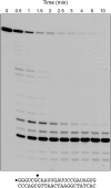
Time course. A reaction mixture containing 25 mM Tris–HCl (pH 7.5), 50 mM NaCl, 5 mM MgCl2, 20 nM 32P-RNA:DNA hybrid duplex (depicted at bottom), and 8 nM RnhA was incubated at 37°C. Aliquots (10 μl) were withdrawn at the times specified and quenched with formamide/EDTA. The reaction products were analyzed by urea–PAGE and visualized by autoradiography.
Figure 4.
Figure 4.
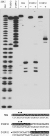
Cleavage of chimeric RNA–DNA junction substrates. Reaction mixtures (10 μl) containing 25 mM Tris–HCl (pH 7.5), 50 mM NaCl, 5 mM MgCl2, 20 nM (200 fmol) 32P-labeled 24-mer duplexes R24, R12D12 or D12R12 (shown at the bottom, with the 32P label denoted by • and the ribonucleotides depicted in white on a black background), and 8 nM (80 fmol) RnhA (where indicated by +) were incubated for 20 min at 37°C. The products were resolved by urea-PAGE and visualized by autoradiography. Alkaline hydrolysis ladders of 32P-labeled R24, R12D12 and D12R12 strand were analyzed in parallel in the three lanes on the left (OH).
Figure 5.
Figure 5.
Minimum RNA requirement. (A) Substrates and cleavage sites. The 32P label is denoted by •. Ribonucleotides are depicted in white on a black background. Principal sites of incision by M. smegmatis RnhA are indicated by black arrowheads. (B) Reaction mixtures (10 μl) containing 25 mM Tris–HCl (pH 7.5), 50 mM NaCl, 5 mM MgCl2, 20 nM (200 fmol) duplex substrate as specified, and either no enzyme (lanes –) or 8 nM (80 fmol) RnhA were incubated at 37°C for 20 min. The products were analyzed by urea-PAGE and visualize by autoradiography. Alkaline hydrolysis ladders of the 32P-labeled strands were analyzed in parallel in lanes OH.
Figure 6.
Figure 6.
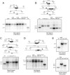
Genotyping of Δrnh mutants. For each panel, schematic representations of the wild type and Δrnh genetic loci are shown with probe location (marked by a grey box ‘P’) and restriction endonuclease sites. Predicted hybridization products are indicated for wild type and deletion alleles. Below these schematics are autoradiograms of Southern blots of restriction endonuclease-digested chromosomal DNA hybridized with the indicated radiolabelled DNA probe from candidate recombinant strains in either wild type or Δrnh backgrounds, as indicated above each lane, along with chromosomal DNA derived from wild type M. smegmatis (WT). (A) Deletion of rnhB in WT (strain Mgm4085), ΔrnhA (strain Mgm4088), ΔrnhD (strain Mgm4090) and ΔrnhA ΔrnhD (strain Mgm4091). (B) Deletion of rnhA in WT (strain Mgm4087), ΔrnhB (strain Mgm4088), and ΔrnhB ΔrnhD (strain Mgm4091). (C) Deletion of rnhC in WT background after removal of the hygR marker via Cre recombinase (strain Mgm4084). (D) Deletion of rnhD in WT (strain Mgm4089), ΔrnhB (strain Mgm4090), ΔrnhA ΔrnhB (strain Mgm4091). (E) Confirmation of the ΔrnhB ΔrnhC double mutant (Mgm4086). Deletion of rnhB in the ΔrnhC background (top panel, see A for schematic) and rnhC in the ΔrnhB background (see C for schematic). In both top and bottom panels, the single mutants ΔrnhB (strain Mgm4085) and ΔrnhC::loxP (strain Mgm4084) were used as the controls in the last lanes, respectively.
Figure 7.
Figure 7.
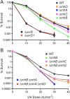
Deletion of rnhB sensitizes M. smegmatis to UV irradiation in stationary phase. (A and B) Strains of the indicated genotypes were grown to stationary phase and exposed to escalating doses of UV irradiation as described in Materials and Methods. Strain survival was quantified by culturing serial 10-fold dilutions of exposed bacteria and normalizing to an unexposed sample from the same culture. When error bars are not visible they are within the plotted symbol.
Figure 8.
Figure 8.
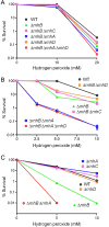
RnhB, aided by RnhA, protects against killing by hydrogen peroxide in stationary phase. Strains of the indicated genotypes either in logarithmic phase (A) or stationary phase (B and C) were exposed to escalating doses of hydrogen peroxide (indicated on the x-axis) for 2 h as described in Materials and Methods. Survival was quantified by culturing serial 10-fold dilutions of exposed bacteria and normalizing to an unexposed sample from the same culture. When error bars are not visible they are within the plotted symbol.
Figure 9.
Figure 9.
Genetic testing of the essentiality of RNase H1 endonuclease activity. A schematic representation of the chromosomal attB locus in strain Mgm4083, which has chromosomal deletions of rnhC and rnhA and a copy of rnhC at the attB locus on an integrated plasmid element conferring kanamycin resistance. Because of spontaneous excision of attB integrated plasmids, replacement with a transfected plasmid carrying a streptomycin resistance gene can be efficiently achieved, allowing swapping of the wild type rnhC gene for wild type and mutated versions of rnhC or rnhA. The results of these marker exchange experiments are given in the table as: (i) the number of strepR colonies recovered after transfection with 50 ng of the plasmid carrying the indicated allele and (ii) the results of genotyping of survivors to confirm allelic exchange.

Similar articles

Cited by

References

    1. Schroeder J.W., Randall J.R., Mattews L.A., Simmons L.A. Ribonucleotides in bacterial DNA. Crit. Rev. Biochem. Mol. Biol. 2014;50:181–193. - PMC - PubMed
    1. Zhu H., Bhattarai H., Yan H., Shuman S., Glickman M. Characterization of Mycobacterium smegmatis PolD2 and PolD1 as RNA/DNA polymerases homologous to the POL domain of bacterial DNA ligase D. Biochemistry. 2012;51:10147–10158. - PMC - PubMed
    1. Ordonez H., Uson M.L., Shuman S. Characterization of three Mycobacterial DinB (DNA polymerase IV) paralogs highlights DinB2 as naturally adept at ribonucleotide incorporation. Nucleic Acids Res. 2014;42:11056–11070. - PMC - PubMed
    1. Ordonez H., Shuman S. Mycobacterium smegmatis DinB2 misincorporates deoxyribonucleotides and ribonucleotides during templated synthesis and lesion bypass. Nucleic Acids Res. 2014;42:12722–12734. - PMC - PubMed
    1. Sharma A., Nair D.T. MsDpo4—a DinB homolog from Mycobacterium smegmatis—is an error-prone DNA polymerase than can promote G;T and T:G mismatches. J. Nucleic Acids. 2012;2012:285481. - PMC - PubMed

MeSH terms

LinkOut - more resources