. 2017 Jan 9;45(1):470-481.
doi: 10.1093/nar/gkw1124. Epub 2016 Nov 29.

Structures of human SRP72 complexes provide insights into SRP RNA remodeling and ribosome interaction

Affiliations

Structures of human SRP72 complexes provide insights into SRP RNA remodeling and ribosome interaction

Matthias M M Becker et al. Nucleic Acids Res. .

Abstract

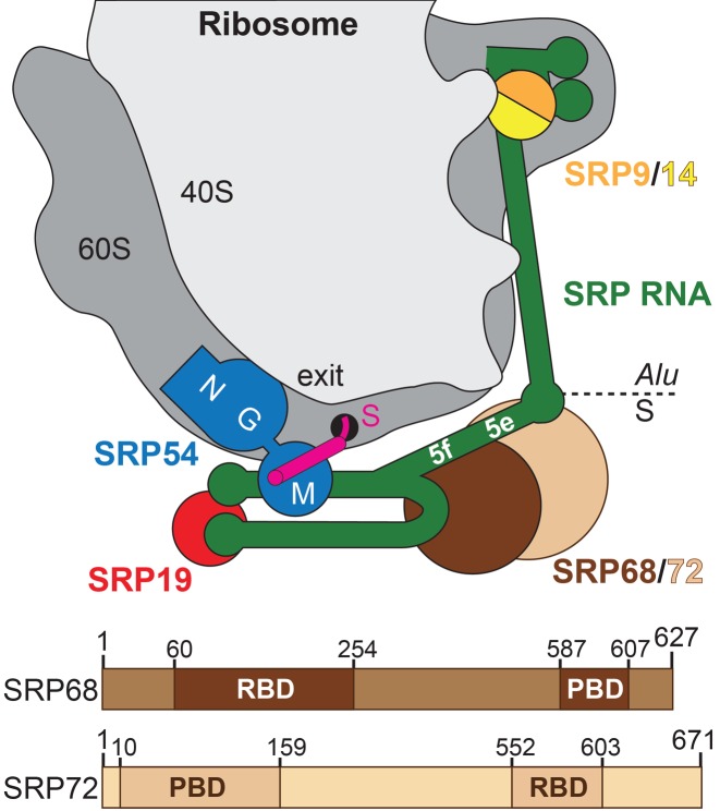
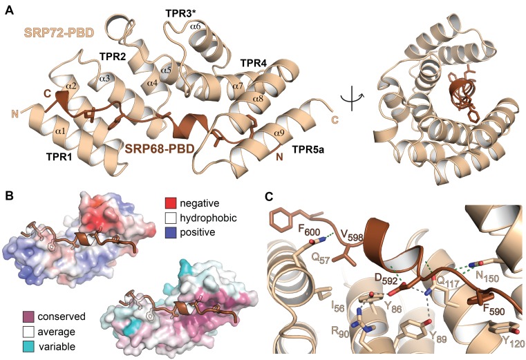
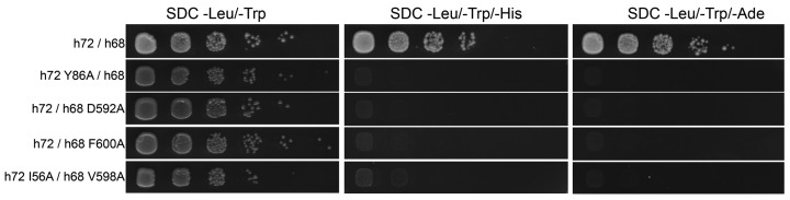
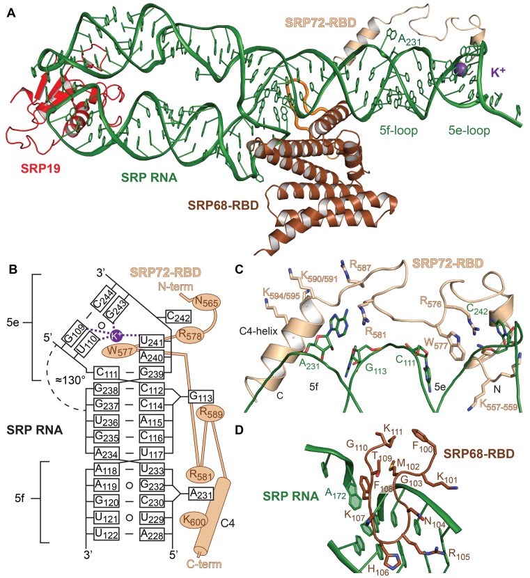
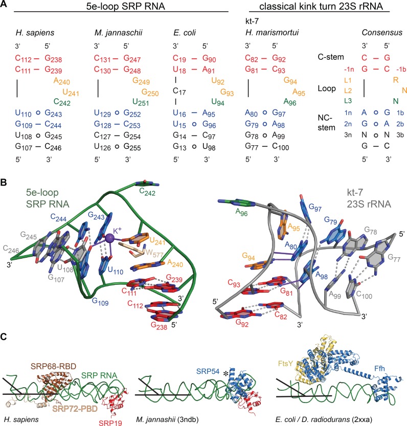
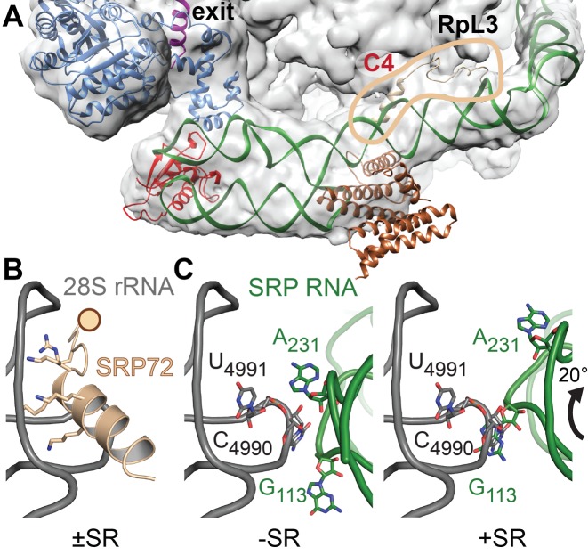
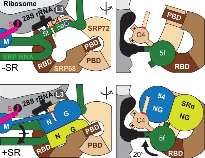
Co-translational protein targeting and membrane protein insertion is a fundamental process and depends on the signal recognition particle (SRP). In mammals, SRP is composed of the SRP RNA crucial for SRP assembly and function and six proteins. The two largest proteins SRP68 and SRP72 form a heterodimer and bind to a regulatory site of the SRP RNA. Despite their essential roles in the SRP pathway, structural information has been available only for the SRP68 RNA-binding domain (RBD). Here we present the crystal structures of the SRP68 protein-binding domain (PBD) in complex with SRP72-PBD and of the SRP72-RBD bound to the SRP S domain (SRP RNA, SRP19 and SRP68) detailing all interactions of SRP72 within SRP. The SRP72-PBD is a tetratricopeptide repeat, which binds an extended linear motif of SRP68 with high affinity. The SRP72-RBD is a flexible peptide crawling along the 5e- and 5f-loops of SRP RNA. A conserved tryptophan inserts into the 5e-loop forming a novel type of RNA kink-turn stabilized by a potassium ion, which we define as K+-turn. In addition, SRP72-RBD remodels the 5f-loop involved in ribosome binding and visualizes SRP RNA plasticity. Docking of the S domain structure into cryo-electron microscopy density maps reveals multiple contact sites between SRP68/72 and the ribosome, and explains the role of SRP72 in the SRP pathway.


Figures

Figure 1.
Figure 1.
SRP68/72 in context of the SRP-RNC. Upper panel: Scheme of human SRP bound to a ribosome—nascent chain complex (gray). Exit: polypeptide tunnel exit; S: signal sequence (magenta); NGM: domains of SRP54 (blue); 5e/5f: loops of SRP RNA (green); SRP9/14 (orange/yellow); SRP19 (red); SRP68 (brown); and SRP72 (sand). Lower panel: Domain architecture of human SRP68/72 (RBD: RNA-binding domain; PBD: protein-binding domain).
Figure 2.
Figure 2.
Structure of the human SRP68/72-PBD complex. (A) Structure of the SRP68/72-PBD complex in side and front views of the solenoid with labeled TPRs and α-helices. Hydrophobic sidechains of SRP68-PBD are shown. (B) Electrostatic surface potential map (top, ±5 kBT/e) and sequence conservation (bottom) of SRP72-PBD. The SRP68-binding groove is mainly hydrophobic and the interface is highly conserved. (C) Details of the interface with the hydrogen-bonding network around D592 including the ‘amide ladder’.
Figure 3.
Figure 3.
Yeast two-hybrid (Y2H) analysis of the SRP68/72-PBD complex. The Y2H is tested in both combinations of activating domain (AD) and binding domain (BD), but only BD-SRP72 and AD-SRP68 are shown. In the first panel, the interaction of the wild-type SRP72-PBD with SRP68-PBD shows the binding of the two domains. Single mutations Y86A of SRP72 (second panel), D592A or F600A of SRP68 (third and fourth panel), as well as the double mutation I56A(SRP72)—V598A(SRP68) (fifth panel) lead to a complete loss of binding. The Y2H of the inverted AD/BD combination is shown in Supplementary Figure S6B.
Figure 4.
Figure 4.
Structure of the human SRP S domain. (A) The quaternary SRP S domain complex. SRP72-RBD (sand) crawls along the 5e- and 5f-loops of SRP RNA (green). A potassium ion (purple sphere) is coordinated in the 5e-loop. The 5f-loop is indicated by the bulged-out adenine A231. The extended loop of SRP68-RBD (brown) in the RNA three-way junction is indicated in orange. (B) Scheme of SRP72-RBD binding to SRP RNA. Non-Watson Crick base pairs are indicated by open circles. (C) Detailed view of SRP72-RBD and its binding to SRP RNA. Residues R576 and W577 are clamped between nucleotides C111 and C242. Adenine A231 stacks onto R581. The lysine-rich region (K557-K559) binds to the 5e-loop and the C4-helix aligns with the 5f-loop. (D) The extended loop of SRP68-RBD (100-111) forms a β-hairpin motif and binds mainly to the backside of the 5f-loop and the SRP RNA three-way junction. The interaction includes a π-stacking between phenylalanine F108 and adenosine A172.
Figure 5.
Figure 5.
Comparison of K-turns with SRP RNA 5e-loops. (A) The consensus sequence for classical K-turns, the K-turn kt-7 of Haloarcula marismortui and SRP RNA 5e-loops of representative members of all three kingdoms of life are shown. In the classical K-turn, the C-stem is formed by two G-C basepairs (red) and lies on the 5′ end of the loop composed of three bulged-out nucleotides (orange, green). Following on the 3′ site a conserved GA/AG (blue) pair forms the NC-stem, which is the most structurally conserved motif in K-turns and which is absent in the 5e-loop of the SRP RNAs. (B) Zoom into the human 5e-loop (green) and and the classical K-turn kt-7 of H. marismortui (54) (PDB 1jj2, gray). The conserved tryptophan W577 (sand) inserts into the 5e-loop and hydrogen bonds are formed in presence of a potassium ion between L2, 1b, 1n and 2n (purple). In the classical K-turn, the two adenines of the NC-stem form hydrogen bonds (purple) with the C-stem (-1n and L1) leading to a kink of approximately 120°. In contrast the SRP RNA 5e-loop kinks to the diametral opposite side of the RNA with approximately 50°. In both cases L3 (green) is bulged-out completely. (C) SRP RNA kinking at the 5e-loop region is present in all kingdoms of life (PDB 2xxa and 3ndb) (55,56). The similar kinking is highlighted by black lines. The kink provides an essential docking platform for the activated targeting complex consisting of the SRP GTPases Ffh (SRP54) and FtsY (SRα). The flexible NG domain (indicated by *) in 3ndb was removed for clarity.
Figure 6.
Figure 6.
SRP–RNC interactions at the C4-contact. (A) Fit of the quaternary S domain complex into the cryo-EM density of a mammalian SRP–RNC complex (EMD-3037 (29)). SRP72-RBD, highlighted by the circle in sand, tethers the S domain on the ribosome (28S rRNA and protein RpL3 at the C4-contact. Large parts of SRP68/72 including the PBDs are flexible and therefore, without density. Color coding as in Figure 1. (B) The interaction of the SRP72-RBD C4-helix with the 3′-terminal three-way junction of 28S rRNA (gray) based on cryo-EM data for SRP/SR-RNC (58) and the human ribosome (PDB 4ugo). The rotation of SRP on the ribosome upon SR interaction requires flexible tethering of the ribosome-bound C4-helix (indicated by a sphere). (C) Rotation of SRP RNA induced by SR binding coincides with a switch of ribosomal contacts from the 5f-loop A231 (left panel) to G113 (right panel). Putative ribosomal base-pairing partners are indicated (C4990 and U4991). In the SR bound state the 5f-loop is exposed, ready to activate the targeting complex. SRP72 is omitted for clarity.
Figure 7.
Figure 7.
Model for SRP72 function in the SRP/SR-RNC complex interface. Scheme for the interaction and repositioning of SRP in context of the RNC±SR (in orthogonal views). The 5e-loop is kinked (potassium ion shown as purple sphere) and forms the docking platform for the targeting complex. The targeting complex is embedded by the SRP68/72 solenoids. The rotation of SRP places the 5f-loop in the targeting complex interface. The C-terminal tail of SRP72 (dashed lines indicate intrinsic disorder, red line indicates caspase cleavage site) locates to the same region and may interact with the targeting complex.

References

    1. Elvekrog M.M., Walter P. Dynamics of co-translational protein targeting. Curr. Opin. Chem. Biol. 2015;29:79–86. - PMC - PubMed
    1. Nyathi Y., Wilkinson B.M., Pool M.R. Co-translational targeting and translocation of proteins to the endoplasmic reticulum. Biochim. Biophys. Acta Mol. Cell Res. 2013;1833:2392–2402. - PubMed
    1. Voorhees R.M., Hegde R.S. Toward a structural understanding of co-translational protein translocation. Curr. Opin. Cell Biol. 2016;41:91–99. - PubMed
    1. Egea P.F., Shan S.-O., Napetschnig J., Savage D.F., Walter P., Stroud R.M. Substrate twinning activates the signal recognition particle and its receptor. Nature. 2004;427:215–221. - PubMed
    1. Focia P.J., Shepotinovskaya I., Seidler J., Freymann D.M. Heterodimeric GTPase core of the SRP targeting complex. Science. 2004;303:373–377. - PMC - PubMed

MeSH terms