. 2017 Mar;22(3):262-273.
doi: 10.1177/1087057116680151. Epub 2016 Dec 13.

High-Throughput Spectral and Lifetime-Based FRET Screening in Living Cells to Identify Small-Molecule Effectors of SERCA

Affiliations

High-Throughput Spectral and Lifetime-Based FRET Screening in Living Cells to Identify Small-Molecule Effectors of SERCA

Tory M Schaaf et al. SLAS Discov. 2017 Mar.

Abstract

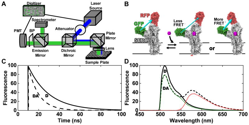
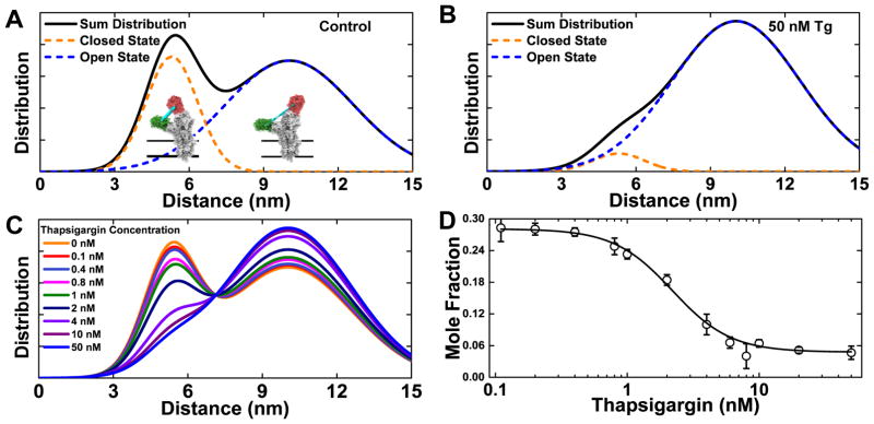
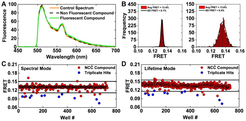
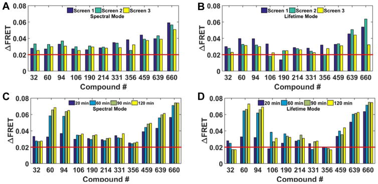
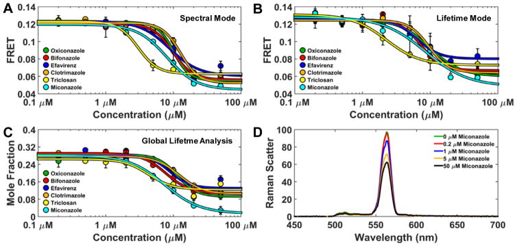
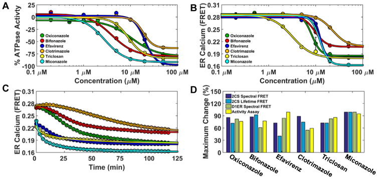
A robust high-throughput screening (HTS) strategy has been developed to discover small-molecule effectors targeting the sarco/endoplasmic reticulum calcium ATPase (SERCA), based on a fluorescence microplate reader that records both the nanosecond decay waveform (lifetime mode) and the complete emission spectrum (spectral mode), with high precision and speed. This spectral unmixing plate reader (SUPR) was used to screen libraries of small molecules with a fluorescence resonance energy transfer (FRET) biosensor expressed in living cells. Ligand binding was detected by FRET associated with structural rearrangements of green fluorescent protein (GFP, donor) and red fluorescent protein (RFP, acceptor) fused to the cardiac-specific SERCA2a isoform. The results demonstrate accurate quantitation of FRET along with high precision of hit identification. Fluorescence lifetime analysis resolved SERCA's distinct structural states, providing a method to classify small-molecule chemotypes on the basis of their structural effect on the target. The spectral analysis was also applied to flag interference by fluorescent compounds. FRET hits were further evaluated for functional effects on SERCA's ATPase activity via both a coupled-enzyme assay and a FRET-based calcium sensor. Concentration-response curves indicated excellent correlation between FRET and function. These complementary spectral and lifetime FRET detection methods offer an attractive combination of precision, speed, and resolution for HTS.

Keywords: biosensor; drug screening; fluorescence lifetime; spectral unmixing; time-resolved FRET.


Conflict of interest statement

Declaration of Conflicting Interests

Dr. Thomas holds equity in and serves as an executive officer for Photonic Pharma LLC. This relationship has been reviewed and managed by the University of Minnesota.

Figures

Figure 1
Figure 1
(A) Diagram of the instrument. (B) The two-color SERCA (2CS) intramolecular FRET biosensor.,, As depicted, the FRET efficiency is dependent on the structural status of SERCA’s domains, which may be affected by the binding of small molecules. Conceptual data (C, lifetime mode) and (D, spectral mode) illustrate the dependence of fluorescence signals on FRET. Solid black curve (D): donor only (no FRET). Dashed black curve (DA): donor plus acceptor (FRET). In (D) dotted curves show the resolution of the spectrum into components corresponding to donor (GFP) and acceptor (RFP) emission.
Figure 2
Figure 2
Lifetime analysis of time-resolved fluorescence decay waveforms resolves structural states of the 2CS biosensor. (A) Two structural states of SERCA are resolved, corresponding to two Gaussian interprobe distance distributions, consistent with a two-state structural model with an equilibrium between closed (5.5 nm FRET distance, orange) and open (10.2 nm FRET distance, blue) structural states. (B) The addition of a saturating dose (50 nM) of the known inhibitor thapsigargin (Tg) shifts this equilibrium substantially toward the open state. (C) Concentration dependence of Tg effect on the structural distribution (n= 8 wells for each concentration). (D) Plot, based on (C), of closed state mole fraction vs Tg.
Figure 3
Figure 3
(A) Fluorescence emission spectra were used to identify and flag potential interference from fluorescent compounds by assessing the similarity index (Eq. 1) of each well from a pilot screen of the NCC1 & 2 small-molecule libraries. A control spectrum (%v/v DMSO well) and non-fluorescent (compound not identified as a FRET hit during screening with 2CS) have a high degree of similarity, as shown as direct overlap of orange and black spectrum. A slightly fluorescent compound is depicted by the green spectrum and was flagged as a potential false-positive hit. The fluorescent profile of all 1152 wells from one NCC screen was assessed using a stringent similarity index threshold; 44 compounds were flagged as potential false-positives due to interference from compound fluorescence. (B) Histogram plots of the wells from one NCC screen after removing potential fluorescent compounds. Gaussian fits depict an increase in precision from spectral mode (left) in comparison to lifetime mode (right), shown as the frequency of FRET efficiency determined by either method and a narrower distribution from spectral mode (average FRET calculated by spectral unmixing or lifetime and the standard deviation determined from the Gaussian fit). (C) One 2CS pilot NCC screen (spectral mode) is shown with a hit threshold set at a 0.02 change in 2CS FRET (4 SD). 16 FRET hits were identified in this screen. 11 of these 2CS FRET hits were found to be reproducible across three independent screens (blue). (D) The same 384-well plates were scanned in lifetime mode. 16 hits were identified using the same threshold set at a 0.02 change in 2CS FRET efficiency (3 SD). In this screen, nine of the reproducible 2CS FRET hits identified in spectral mode were also FRET hits as assessed by lifetime mode (blue).
Figure 4
Figure 4
Reproducible FRET hits assessed across independent screens and time course studies. (A) Spectral mode identified eleven reproducible 2CS FRET hits using a threshold of 0.02 change in FRET (red line). The reproducibility of each 2CS FRET hit, after 20 min incubation, across three independent screens is shown. The ΔFRET from each compound remains consistent from screen to screen. ΔFRET was calculated by assessing the change in FRET of 2CS from the average FRET, determined by the Gaussian fit of all wells not flagged as fluorescence compounds. (B) Lifetime mode assessment of the eleven reproducible spectral FRET hits. Nine of the eleven FRET hits were reproducible (triplicate) hits using a 0.02 FRET threshold. Compounds #106 and190 were not identified as lifetime FRET hits in one of the three independent screens. (C) Spectral FRET hits evaluated by time-course studies. Each independent pilot screen was scanned in spectral mode after 20, 60, 90, and 120 minutes of compound incubation. The change in 2CS FRET efficiency (ΔFRET) of each compound is plotted and each reproducible FRET hit, identified as a hit using the spectral unmixing method, remained a hit over multiple time points. The 2CS FRET hits depicted here were from screen 2 (turquoise bars in A and B). Compounds #60, 94, 459, 639, and 660 exhibited an increased FRET change over time. (D) Lifetime FRET change evaluated by time-course studies. Ten compounds from screen 2 were identified as hits, after 20 minutes of compound incubation, using a threshold set at 0.02 FRET change (red bar). Compounds #32 and 356 displayed a modest reduction in 2CS ΔFRET at the later time points. Compounds # 60, 94, 459, 639, and 660 again exhibited an increased FRET change over time.
Figure 5
Figure 5
Multi-parameter concentration-dependent effect of FRET hits (A) Spectral mode analysis of the reproducible 2CS FRET hits. Compounds were dispensed into 384 well plates, across an eight-point concentration-gradient (n=4 for each concentration). Six representative compounds produced a dose-dependent FRET change as evaluated in spectral mode. These compounds altered FRET with micromolar affinities with subtle differences across the compounds. (B) The same 384-well plate was evaluated using lifetime mode and demonstrated excellent agreement in the dose-dependent FRET change across two independent FRET measurements. (C) Global analysis of the lifetime data depicts a dose-dependent change in the mole fraction of the closed 2CS headpiece (5.5 nm distance distribution). Using this distance distribution model, the confirmed reproducible hits perturbed the 2CS structural equilibrium between open and closed states. All of the hits decreased 2CS FRET, indicating increased distance between GFP and RFP. (D) Water Raman spectrum acquired from compound-only wells of the known compound aggregator miconazole demonstrates ultra-high-sensitivity of spectral recording. Compound aggregation dose-dependently causes more light to be absorbed and decreases inelastic light scattering (Raman band).
Figure 6
Figure 6
Functional characterization of FRET hits on SERCA ATPase activity and ER calcium content. (A) 2CS FRET hits inhibit SERCA ATPase activity. NADH-enzyme coupled activity assay of purified SERCA was measured at eight different concentrations of the reproducible FRET hits. The maximal rate of SERCA activity was measured at saturating calcium (10 μM) after 20 minute incubation with compounds and dose-dependent inhibition was observed. (B) Endoplasmic reticulum calcium was depleted by the 2CS FRET hits. ER calcium was monitored in live-cells overexpressing the endoplasmic reticulum localized calcium FRET sensor (D1ER). D1ER FRET is dependent on calcium concentration, where less calcium causes a reduction in FRET. ER calcium levels were monitored over time in response to an eight-point concentration gradient of each hit compound. A 384 well plate was repeatedly scanned (every three minutes) with D1ER cells. The dose-dependent FRET change (ER calcium depletion) after 120 minutes compound incubation is shown and depicts differential depletion at each compound concentration. (C) Maximal ER calcium depletion in the presence of a saturating dose (50 μM) of each compound (decreased D1ER FRET) was assessed over a 120 minute period. The 2CS FRET hits displayed time-dependent and compound-specific ER calcium depletion. Miconazole (turquoise) exhibited both maximal SERCA ATPase Vmax inhibition and the largest amount of ER depletion. (D) Structure and activity assay correlation of 2CS FRET hits. The maximal change (percent change) of the structural FRET change from the 2CS FRET biosensor as well as the maximal change from two different functional assay (ATPase activity assay and D1ER calcium depletion). are shown.

References

    1. Cornea RL, Gruber SJ, Lockamy EL, et al. High-throughput FRET assay yields allosteric SERCA activators. J Biomol Screen. 2013;18:97–107. - PMC - PubMed
    1. Gruber SJ, Cornea RL, Li J, et al. Discovery of enzyme modulators via high-throughput time-resolved FRET in living cells. J Biomol Screen. 2014;19:215–222. - PMC - PubMed
    1. Bleeker NP, Cornea RL, Thomas DD, et al. A Novel SERCA Inhibitor Demonstrates Synergy with Classic SERCA Inhibitors and Targets Multidrug-Resistant AML. Mol Pharm. 2013;10(11):4358–66. - PMC - PubMed
    1. Kang S, Dahl R, Hsieh W, et al. Small Molecular Allosteric Activator of the Sarco/Endoplasmic Reticulum Ca2+-ATPase (SERCA) Attenuates Diabetes and Metabolic Disorders. J Biol Chem. 2015;291(10):5185–98. - PMC - PubMed
    1. Brini M, Carafoli E. Calcium pumps in health and disease. Physiol Rev. 2009;89:1341–1378. - PubMed

Publication types

MeSH terms

Substances