Quantitative label-free mass spectrometry analysis of formalin-fixed, paraffin-embedded tissue representing the invasive cutaneous malignant melanoma proteome
- PMID: 27899996
- PMCID: PMC5103945
- DOI: 10.3892/ol.2016.5101
Quantitative label-free mass spectrometry analysis of formalin-fixed, paraffin-embedded tissue representing the invasive cutaneous malignant melanoma proteome
Abstract
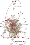
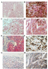
Understanding the events at a protein level that govern the progression from melanoma in situ to invasive melanoma are important areas of current research to be developed. Recent advances in the analysis of formalin-fixed, paraffin-embedded tissue by proteomics, particularly using the filter-aided sample preparation protocol, has opened up the possibility of studying vast archives of clinical material and associated medical records. In the present study, quantitative protein profiling was performed using tandem mass spectrometry, and the proteome differences between melanoma in situ and invasive melanoma were compared. Biological pathway analyses revealed several signalling pathways differing between melanoma in situ and invasive melanoma, including metabolic pathways and the phosphoinositide 3-kinase-Akt signalling pathway. Selected proteins of interest (14-3-3ε and fatty acid synthase) were subsequently investigated using immunohistochemical analysis of tissue microarrays. Identifying the key proteins that play significant roles in the establishment of a more invasive phenotype in melanoma may ultimately aid diagnosis and treatment decisions.
Keywords: 14-3-3ε; FASN; FFPE; mass spectrometry; melanoma; proteomics.
Figures


Similar articles
-
Proteome, phosphoproteome, and N-glycoproteome are quantitatively preserved in formalin-fixed paraffin-embedded tissue and analyzable by high-resolution mass spectrometry.J Proteome Res. 2010 Jul 2;9(7):3688-700. doi: 10.1021/pr100234w. J Proteome Res. 2010. PMID: 20469934
-
Qualitative and quantitative proteomic analysis of formalin-fixed paraffin-embedded (FFPE) tissue.Methods Mol Biol. 2015;1295:109-15. doi: 10.1007/978-1-4939-2550-6_10. Methods Mol Biol. 2015. PMID: 25820718
-
Label-free protein profiling of formalin-fixed paraffin-embedded (FFPE) heart tissue reveals immediate mitochondrial impairment after ionising radiation.J Proteomics. 2012 Apr 18;75(8):2384-95. doi: 10.1016/j.jprot.2012.02.019. Epub 2012 Feb 23. J Proteomics. 2012. PMID: 22387116
-
Excavation of a buried treasure--DNA, mRNA, miRNA and protein analysis in formalin fixed, paraffin embedded tissues.Histol Histopathol. 2011 Jun;26(6):797-810. doi: 10.14670/HH-26.797. Histol Histopathol. 2011. PMID: 21472693 Review.
-
Update on proteomic studies of formalin-fixed paraffin-embedded tissues.Expert Rev Proteomics. 2019 Jun;16(6):513-520. doi: 10.1080/14789450.2019.1615452. Epub 2019 May 16. Expert Rev Proteomics. 2019. PMID: 31094245 Review.
Cited by
-
14-3-3ε: a protein with complex physiology function but promising therapeutic potential in cancer.Cell Commun Signal. 2024 Jan 26;22(1):72. doi: 10.1186/s12964-023-01420-w. Cell Commun Signal. 2024. PMID: 38279176 Free PMC article. Review.
-
Characterization of long noncoding RNA and messenger RNA signatures in melanoma tumorigenesis and metastasis.PLoS One. 2017 Feb 22;12(2):e0172498. doi: 10.1371/journal.pone.0172498. eCollection 2017. PLoS One. 2017. PMID: 28225791 Free PMC article.
-
Proteomic Profiling of Archived Tissue of Primary Melanoma Identifies Proteins Associated with Metastasis.Int J Mol Sci. 2020 Oct 31;21(21):8160. doi: 10.3390/ijms21218160. Int J Mol Sci. 2020. PMID: 33142795 Free PMC article.
-
Proteome Profiling of Primary Pancreatic Ductal Adenocarcinomas Undergoing Additive Chemoradiation Link ALDH1A1 to Early Local Recurrence and Chemoradiation Resistance.Transl Oncol. 2018 Dec;11(6):1307-1322. doi: 10.1016/j.tranon.2018.08.001. Epub 2018 Aug 30. Transl Oncol. 2018. PMID: 30172883 Free PMC article.
-
Towards Precision Dermatology: Emerging Role of Proteomic Analysis of the Skin.Dermatology. 2022;238(2):185-194. doi: 10.1159/000516764. Epub 2021 Jun 1. Dermatology. 2022. PMID: 34062531 Free PMC article. Review.
References
-
- Masters GA, Krilov L, Bailey HH, Brose MS, Burstein H, Diller LR, Dizon DS, Fine HA, Kalemkerian GP, Moasser M, et al. Clinical cancer advances 2015: Annual report on progress against cancer from the American society of clinical oncology. J Clin Oncol. 2015;33:786–809. doi: 10.1200/JCO.2014.59.9746. - DOI - PubMed
LinkOut - more resources
Full Text Sources
Other Literature Sources
Miscellaneous
