

Surgical treatment of rare giant malignant tumors of the scalp: A report of 3 cases with different tumor types
- PMID: 27900013
- PMCID: PMC5103960
- DOI: 10.3892/ol.2016.5113
Surgical treatment of rare giant malignant tumors of the scalp: A report of 3 cases with different tumor types
Abstract
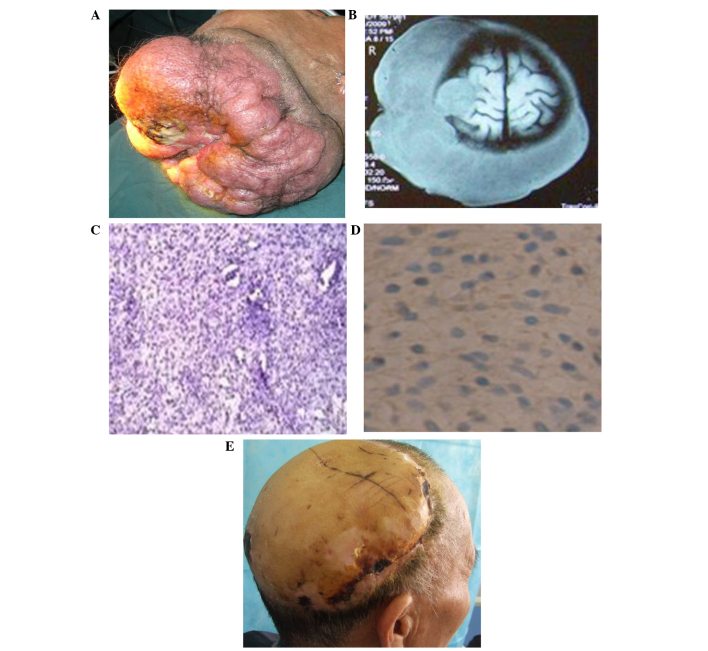
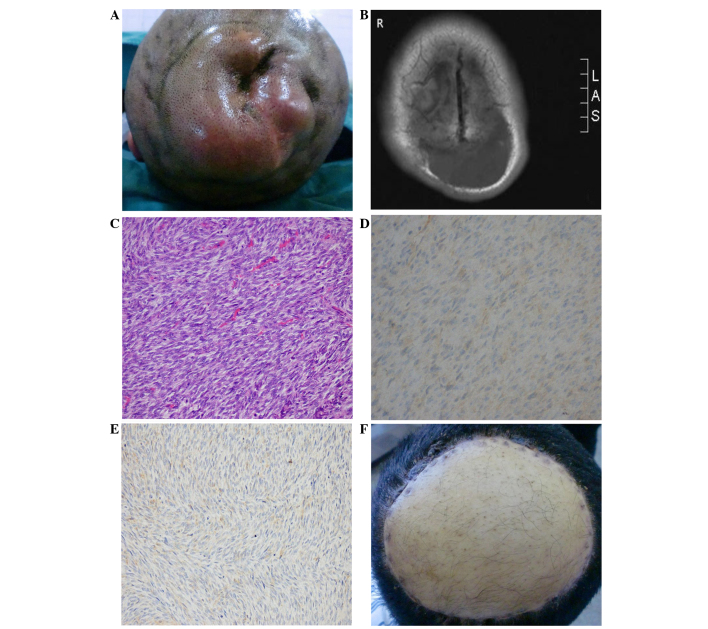
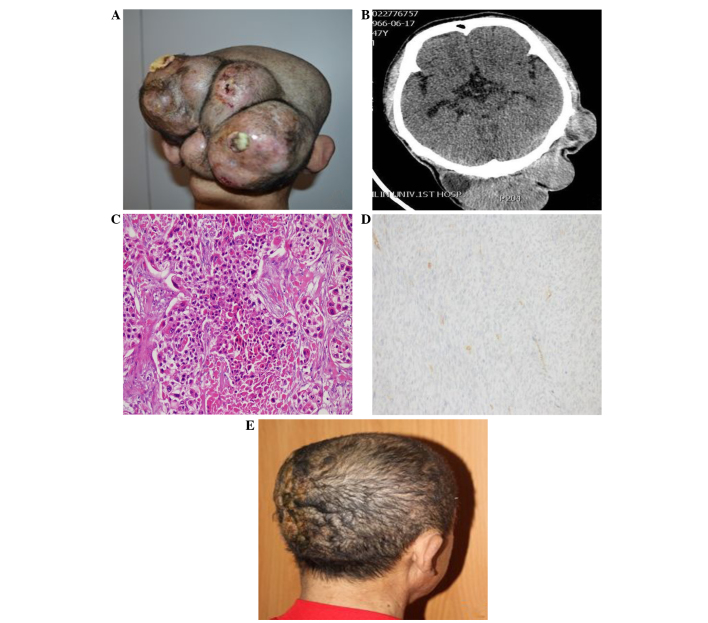
The scalp is the most frequent site of occurrence of malignant tumors. As an area that is generally neglected by the patient and not closely monitored during physical examinations, scalp tumors can go unnoticed until they become malignant. The present study reports 3 cases of rare giant malignant tumors of the scalp, namely a peripheral nerve sheath tumor, a fibrous tumor and a malignant proliferating trichilemmal tumor, that were treated at The First Bethune Hospital of Jilin University (Changchun, China). Vascularized free anterolateral thigh flap surgery was performed in 2 of the 3 cases. A local flap repair was applied to the third case. The implanted skin grafts remained viable post-operatively and wound repair was uneventful. No signs of malignancy were detected on the edge of the pathological section upon closer pathological examination. In the follow-up period, no recurrence was detected in any of the cases.
Keywords: malignant tumors of the scalp; surgical treatment.
Figures
Similar articles
-
Malignant peripheral nerve sheath tumor of the scalp: Two rare case reports.Surg Neurol Int. 2018 May 15;9:102. doi: 10.4103/sni.sni_196_17. eCollection 2018. Surg Neurol Int. 2018. PMID: 29900032 Free PMC article.
-
Giant Malignant Peripheral Nerve Sheath Tumor of the Scalp: Case Report and Review of the Literature.World Neurosurg. 2020 Jun;138:246-252. doi: 10.1016/j.wneu.2020.03.039. Epub 2020 Mar 13. World Neurosurg. 2020. PMID: 32179188 Review.
-
Malignant fibrous histiocytoma of the scalp. Multidisciplinary treatment.J Eur Acad Dermatol Venereol. 1999 Nov;13(3):175-82. J Eur Acad Dermatol Venereol. 1999. PMID: 10642053
-
Malignant proliferating trichilemmal tumor.Am J Clin Oncol. 2001 Aug;24(4):351-3. doi: 10.1097/00000421-200108000-00006. Am J Clin Oncol. 2001. PMID: 11474259
-
Malignant transformation of a giant proliferating trichilemmal tumor of the scalp: patient report and literature review.Ann Plast Surg. 1998 Sep;41(3):314-6. doi: 10.1097/00000637-199809000-00017. Ann Plast Surg. 1998. PMID: 9746092 Review.
Cited by
-
Malignant Peripheral Nerve Sheath Scalp Tumor: A Short-Term Institutional Experience with Literature Review.Asian J Neurosurg. 2023 Mar 27;18(1):157-164. doi: 10.1055/s-0043-1763524. eCollection 2023 Mar. Asian J Neurosurg. 2023. PMID: 37056889 Free PMC article.
-
A Rare Case of a Malignant Proliferating Trichilemmal Tumor: A Molecular Study Harboring Potential Therapeutic Significance and a Review of Literature.Dermatopathology (Basel). 2024 Dec 10;11(4):354-363. doi: 10.3390/dermatopathology11040038. Dermatopathology (Basel). 2024. PMID: 39727620 Free PMC article.
-
Malignant Proliferating Trichilemmal Tumor: A Subtle Presentation in an African American Woman and Review of Immunohistochemical Markers for This Rare Condition.Cureus. 2021 Aug 18;13(8):e17289. doi: 10.7759/cureus.17289. eCollection 2021 Aug. Cureus. 2021. PMID: 34552829 Free PMC article.
-
[Gigantic exophytically growing tumor of the occipital scalp : A complex diagnostic and reconstructive challenge].Chirurg. 2017 Oct;88(10):879-883. doi: 10.1007/s00104-017-0425-0. Chirurg. 2017. PMID: 28421260 German. No abstract available.
-
Association of Flame-Retardant Clothing With Mycosis Fungoides: A Retrospective Analysis.Dermatol Pract Concept. 2022 Apr 1;12(2):e2022091. doi: 10.5826/dpc.1202a91. eCollection 2022 May. Dermatol Pract Concept. 2022. PMID: 35646447 Free PMC article.
References
-
- Richmond HM, Duvic M, Macfarlane DF. Primary and metastatic malignant tumors of the scalp: An update. Am J Clin Dermatol. 2010;11:233–246. - PubMed
-
- Jia X, Yang J. Progress of malignant peripheral nerve sheath tumors study. Int J Orthop. 2014;35:164–166.
-
- Fletcher CDM, Unni KK, Mertens F, editors. Pathology and Genetics of Tumours of Soft Tissue and Bone. IARC Press; Lyon: 2002. World Health Organization Classification of Tumours.
LinkOut - more resources
Full Text Sources
Other Literature Sources
Research Materials