

Integration of genomics and histology revises diagnosis and enables effective therapy of refractory cancer of unknown primary with PDL1 amplification
- PMID: 27900363
- PMCID: PMC5111004
- DOI: 10.1101/mcs.a001180
Integration of genomics and histology revises diagnosis and enables effective therapy of refractory cancer of unknown primary with PDL1 amplification
Abstract
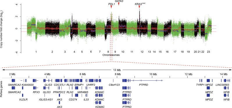
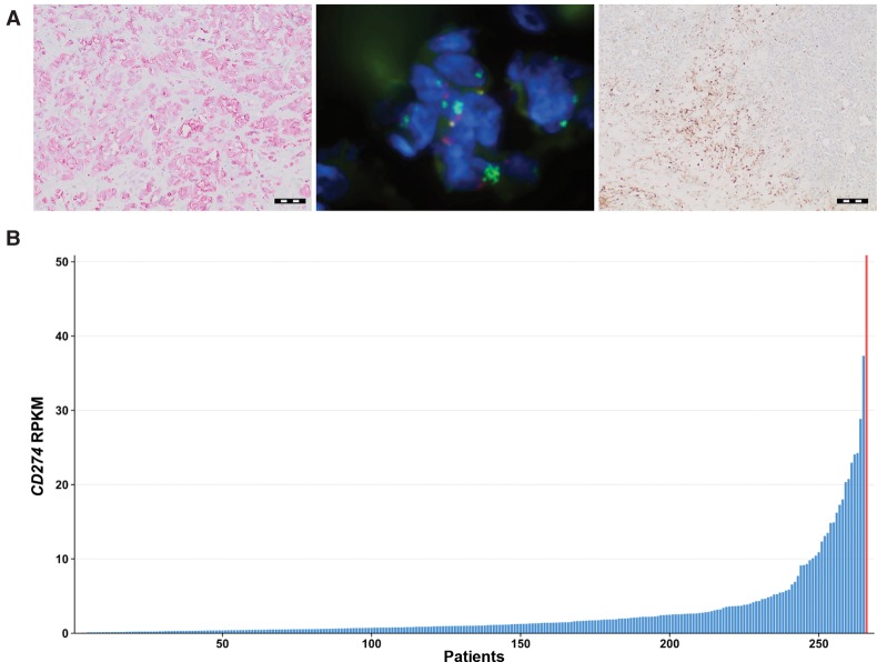
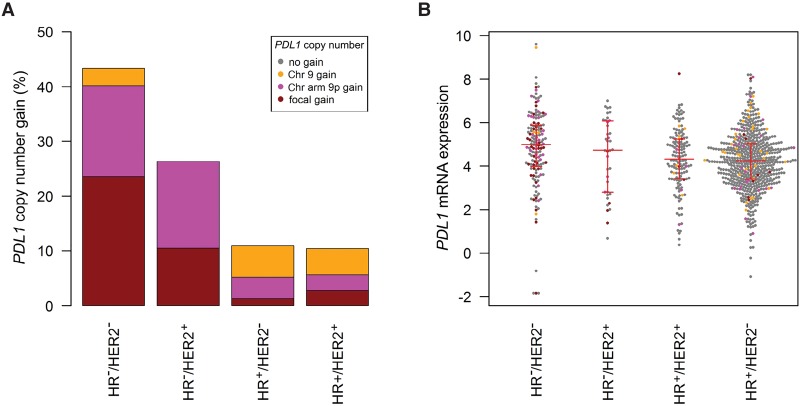
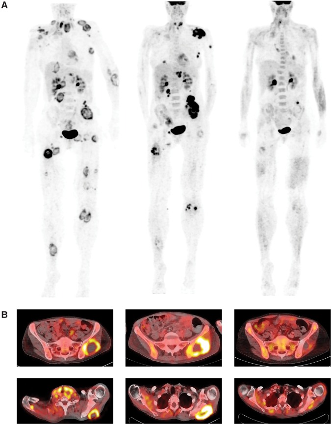
Identification of the tissue of origin in cancer of unknown primary (CUP) poses a diagnostic challenge and is critical for directing site-specific therapy. Currently, clinical decision-making in patients with CUP primarily relies on histopathology and clinical features. Comprehensive molecular profiling has the potential to contribute to diagnostic categorization and, most importantly, guide CUP therapy through identification of actionable lesions. We here report the case of an advanced-stage malignancy initially mimicking poorly differentiated soft-tissue sarcoma that did not respond to multiagent chemotherapy. Molecular profiling within a clinical whole-exome and transcriptome sequencing program revealed a heterozygous, highly amplified KRAS G12S mutation, compound-heterozygous TP53 mutation/deletion, high mutational load, and focal high-level amplification of Chromosomes 9p (including PDL1 [CD274] and JAK2) and 10p (including GATA3). Integrated analysis of molecular data and histopathology provided a rationale for immune checkpoint inhibitor (ICI) therapy with pembrolizumab, which resulted in rapid clinical improvement and a lasting partial remission. Histopathological analyses ruled out sarcoma and established the diagnosis of a poorly differentiated adenocarcinoma. Although neither histopathology nor molecular data were able to pinpoint the tissue of origin, our analyses established several differential diagnoses including triple-negative breast cancer (TNBC). We analyzed 157 TNBC samples from The Cancer Genome Atlas, revealing PDL1 copy number gains coinciding with excessive PDL1 mRNA expression in 24% of cases. Collectively, these results illustrate the impact of multidimensional tumor profiling in cases with nondescript histology and immunophenotype, show the predictive potential of PDL1 amplification for immune checkpoint inhibitors (ICIs), and suggest a targeted therapeutic strategy in Chromosome 9p24.1/PDL1-amplified cancers.
Keywords: multifocal breast carcinoma; neoplasm of the gastrointestinal tract.
Figures
References
-
- Barrett MT, Anderson KS, Lenkiewicz E, Andreozzi M, Cunliffe HE, Klassen CL, Dueck AC, McCullough AE, Reddy SK, Ramanathan RK, et al. 2015. Genomic amplification of 9p24.1 targeting JAK2, PD-L1, and PD-L2 is enriched in high-risk triple negative breast cancer. Oncotarget 6: 26483–26493. - PMC - PubMed
-
- Budczies J, Denkert MBC, Klauschen F, Gröschel S, Darb-Esfahani S, Pfarr N, Leichsenring J, Onozato ML, Lennerz JK, Dietel M, et al. 2016. Pan-cancer analysis of copy number changes in programmed death-ligand 1 (PD-L1, CD274)—associations with gene expression, mutational load and survival. Genes Chromosomes Cancer 55: 626–639. - PubMed
-
- Buisseret L, Specht J, Dees EC, Berger R, Gupta S, Geva R, Pusztai L, Gause CK, Karantza V, Nanda R. 2015. 14P * KEYNOTE-012: a phase Ib study of pembrolizumab (MK-3475) in patients (pts) with metastatic triple-negative breast cancer (mTNBC). Ann Oncol 26suppl 3: ii6–iii6.
Publication types
MeSH terms
Substances
LinkOut - more resources
Full Text Sources
Other Literature Sources
Research Materials
Miscellaneous