Review
. 2017 Jun;34(3):351-361.
doi: 10.1007/s10719-016-9749-0. Epub 2016 Nov 30.

Mast cell glycosaminoglycans

Affiliations
Review

Mast cell glycosaminoglycans

B Mulloy et al. Glycoconj J. 2017 Jun.

Abstract

Mast cells contain granules packed with a mixture of proteins that are released on degranulation. The proteoglycan serglycin carries an array of glycosaminoglycan (GAG) side chains, sometimes heparin, sometimes chondroitin or dermatan sulphate. Tight packing of granule proteins is dependent on the presence of serglycin carrying these GAGs. The GAGs of mast cells were most intensively studied in the 1970s and 1980s, and though something is known about the fine structure of chondroitin sulphate and dermatan sulphate in mast cells, little is understood about the composition of the heparin/heparan sulphate chains. Recent emphasis on the analysis of mast cell heparin from different species and tissues, arising from the use of this GAG in medicine, lead to the question of whether variations within heparin structures between mast cell populations are as significant as variations in the mix of chondroitins and heparins.

Keywords: Chondroitin; Dermatan; Glycosaminoglycan; Heparin; Mast cell; Serglycin.


Conflict of interest statement

Conflict of interest

The authors declare that they have no conflicts of interest.

Ethical approval

This article does not contain any studies with human participants or animals performed by any of the authors.

Figures

Fig. 1
Fig. 1
a, b Transmission electron micrographs showing an intact rat peritoneal MC (a) and a MC undergoing anaphylactic degranulation (b). The block arrows indicate regions where multiple granules have fused; the arrow depicts an exocytosed granule remnant. c, d Scanning electron micrographs showing an intact (c) and degranulated (d) rat peritoneal MC. Note the extensive membrane alterations in d. Arrows in d depict exocytosed granule remnants. Images are courtesy of Prof. Giuliano Zabucchi, Prof. Maria Rosa Soranzo, and Dr. Francesca Vita (Electron Microscopy Section of Centro Coordinamento e Sviluppo Progetti e Apparecchiature (CSPA), University of Trieste). Figure and caption reproduced with permission from [39]
Fig. 2
Fig. 2
GAG structures found in mast cells. a The main repeating unit of heparan sulphate, [ −4)-β-D-GlcA-(1 → 4)-α-D-GlcNAc-(1- ] (GlcA-GlcNAc). This structure forms the precursor polysaccharide, which is transformed by a series of enzymes into b the main repeating unit of heparin [−4)-α-L-IdoA(2SO3 )-(1 → 4)-α-D-GlcNSO3 (6SO3 )-(1-] (IdoA2S-GlcNS6S). c the pentasaccharide with high affinity for antithrombin, as found in porcine mucosal heparin; essential substituents are marked with an asterisk. The enzyme 3-O-sulfotransferase (3-OST) isoform 1 adds the unusual 3-O-sulphate substituent (indicated with a double asterisk) to the central N-, 6-disulphated glucosamine in the GlNR6S-GlcA-GlcNS,6S–IdoA2S-GlcNS,6S sequence
Fig. 3
Fig. 3
The conversion of the chondroitin backbone to CS-A, CS-B, CS-C, CS-D, CS-E and CS-diB by the enzymes chondroitin-4-sulfotranferase (C4ST), GlcA 5-epimerase (DS-epi), dermatan 4-O-sulfotransferase (D4ST), chondroitin 6-sulfotransferase (CS6ST) uronosyl 2-O-sulfotransferase (UST) GalNAc 4-O- sulphate 6-O-sulfotransferase (GalNAc4S-6ST). Reproduced with permission from [15]
Fig. 4
Fig. 4
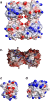
Heparin interactions with proteases a A β-tryptase tetramer crystal structure (1A0L.pdb), coloured by charge where blue is positive and red negative. This view clearly shows the restricted central active site. Clusters of basic residues, coloured blue, might accommodate a heparin chain binding to two monomers and so stabilising the active tetramer. Diagram made in Discovery Studio Visualiser, Accelrys Software Inc. b An α-tryptase tetramer crystal structure (1LTO.pdb), turned over by 90° to show the equivalent of the top face as compared with A). This view shows an almost continuous line of basic amino acid residues aligning with a long heparin molecule, shown in ball and stick format. Reproduced from [50] with permission. c A chymase monomer crystal structure (4KP0.pdb), showing clusters of basic amino acid residues (blue) on opposite sides of the charge-coloured surface, with a cluster of acidic residues (red) between them. The orientation of these two basic patches on the surface of chymase might allow this enzyme to pack between heparin chains of serglycin. The active site groove is on the left face of this diagram. Diagram made in Discovery Studio Visualiser, Accelrys Software Inc. d A granzyme B monomer crystal structure (1IAU.pdb) showing a substantial cluster of basic residues (blue) at the top of the charge-coloured surface. The active site is on the left face of the diagram. Diagram made in Discovery Studio Visualiser, Accelrys Software Inc

Similar articles

Cited by

References

    1. Crivellato E, Ribatti D. The mast cell: an evolutionary perspective. Biol. Rev. Camb. Philos. Soc. 2010;85:347–360. doi: 10.1111/j.1469-185X.2009.00105.x. - DOI - PubMed
    1. Wernersson S, Pejler G. Mast cell secretory granules: armed for battle. Nat. Rev. Immunol. 2014;14:478–494. doi: 10.1038/nri3690. - DOI - PubMed
    1. Ribatti D. The development of human mast cells. An historical reappraisal. Exp. Cell Res. 2016;342:210–215. doi: 10.1016/j.yexcr.2016.03.013. - DOI - PubMed
    1. Ronnberg E, Melo FR, Pejler G. Mast cell proteoglycans. J. Histochem. Cytochem. 2012;60:950–962. doi: 10.1369/0022155412458927. - DOI - PMC - PubMed
    1. Esko, J. D., Kimata, K., & Lindahl, U. 2009, Proteoglycans and Sulfated Glycosaminoglycans. In: Varki A. et al., eds. Essentials of Glycobiology, 2 ed. Pp. 219–248, Cold Spring Harbor Press (2009). - PubMed

MeSH terms

LinkOut - more resources