Is There Asymmetry Between the Concave and Convex Pedicles in Adolescent Idiopathic Scoliosis? A CT Investigation
- PMID: 27900714
- PMCID: PMC5289204
- DOI: 10.1007/s11999-016-5188-2
Is There Asymmetry Between the Concave and Convex Pedicles in Adolescent Idiopathic Scoliosis? A CT Investigation
Abstract
Background: Adolescent idiopathic scoliosis is a complex three-dimensional deformity of the spine characterized by deformities in the sagittal, coronal, and axial planes. Spinal fusion using pedicle screw instrumentation is a widely used method for surgical correction in severe (coronal deformity, Cobb angle > 45°) adolescent idiopathic scoliosis curves. Understanding the anatomic difference in the pedicles of patients with adolescent idiopathic scoliosis is essential to reduce the risk of neurovascular or visceral injury through pedicle screw misplacement.
Questions/purposes: To use CT scans (1) to analyze pedicle anatomy in the adolescent thoracic scoliotic spine comparing concave and convex pedicles and (2) to assess the intra- and interobserver reliability of these measurements to provide critical information to spine surgeons regarding size, length, and angle of projection.
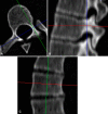
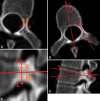
Methods: Between 2007 and 2009, 27 patients with adolescent idiopathic scoliosis underwent thoracoscopic anterior correction surgery by two experienced spinal surgeons. Preoperatively, each patient underwent a CT scan as was their standard of care at that time. Twenty-two patients (mean age, 15.7 years; SD, 2.4 years; range, 11.6-22 years) (mean Cobb angle, 53°; SD, 5.3°; range, 42°-63°) were selected. Inclusion criteria were a clinical diagnosis of adolescent idiopathic scoliosis, female, and Lenke type 1 adolescent idiopathic scoliosis with the major curve confined to the thoracic spine. Using three-dimensional image analysis software, the pedicle width, inner cortical pedicle width, pedicle height, inner cortical pedicle height, pedicle length, chord length, transverse pedicle angle, and sagittal pedicle angles were measured. Randomly selected scans were remeasured by two of the authors and the reproducibility of the measurement definitions was validated through limit of agreement analysis.
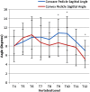
Results: The concave pedicle widths were smaller compared with the convex pedicle widths at T7, T8, and T9 by 37% (3.44 mm ± 1.16 mm vs 4.72 mm ± 1.02 mm; p < 0.001; mean difference, 1.27 mm; 95% CI, 0.92 mm-1.62 mm), 32% (3.66 mm ± 1.00 mm vs 4.82 mm ± 1.10 mm; p < 0.001; mean difference, 1.16 mm; 95% CI, 0.84 mm-1.49 mm), and 25% (4.10 mm ± 1.57 mm vs 5.12 mm ± 1.17 mm; p < 0.001; mean difference, 1.02 mm; 95% CI, 0.66 mm-1.39 mm), respectively. The concave pedicle heights were smaller than the convex at T5 (9.43 mm ± 0.98 vs 10.63 mm ± 1.10 mm; p = 0.002; mean difference, 1.02 mm; 95% CI, 0.59 mm-1.45 mm), T6 (8.87 mm ± 1.37 mm vs 10.88 mm ± 0.81 mm; p < 0.001; mean difference, 2.02 mm; 95% CI, 1.40 mm-2.63 mm), T7 (9.09 mm ± 1.24 mm vs 11.35 mm ± 0.84 mm; p < 0.001; mean difference, 2.26 mm; 95% CI, 1.81 mm-2.72 mm), and T8 (10.11 mm ± 1.05 mm vs 11.86 mm ± 0.88 mm; p < 0.001; mean difference, 1.75 mm; 95% CI, 1.30 mm-2.19 mm). Conversely, the concave transverse pedicle angle was larger than the convex at levels T6 (11.37° ± 4.48° vs 8.82° ± 4.31°; p = 0.004; mean difference, 2.54°; 95% CI, 1.10°-3.99°), T7 (12.69° ± 5.93° vs 8.65° ± 3.79°; p = 0.002; mean difference, 4.04°; 95% CI, 1.90°-6.17°), T8 (13.24° ± 5.28° vs 7.66° ± 4.87°; p < 0.001; mean difference, 5.58°; 95% CI, 2.99°-8.17°), and T9 (19.95° ± 5.69° vs 8.21° ± 4.02°; p < 0.001; mean difference, 4.74°; 95% CI, 2.68°-6.80°), indicating a more posterolateral to anteromedial pedicle orientation.
Conclusions: There is clinically important asymmetry in the morphologic features of pedicles in individuals with adolescent idiopathic scoliosis. The concave side of the curve compared with the convex side is smaller in height and width periapically. Furthermore, the trajectory of the pedicle is more acute on the convex side of the curve compared with the concave side around the apex of the curve. Knowledge of these anatomic variations is essential when performing scoliosis correction surgery to assist with selecting the correct pedicle screw size and trajectory of insertion to reduce the risk of pedicle wall perforation and neurovascular injury.
Figures


Similar articles
-
What is the Difference in Morphologic Features of the Thoracic Pedicle Between Patients With Adolescent Idiopathic Scoliosis and Healthy Subjects? A CT-based Case-control Study.Clin Orthop Relat Res. 2017 Nov;475(11):2765-2774. doi: 10.1007/s11999-017-5448-9. Epub 2017 Aug 1. Clin Orthop Relat Res. 2017. PMID: 28766159 Free PMC article.
-
Relationship between vertebral morphology and the potential risk of spinal cord injury by pedicle screw in adolescent idiopathic scoliosis.Clin Neurol Neurosurg. 2018 Sep;172:143-150. doi: 10.1016/j.clineuro.2018.07.007. Epub 2018 Jul 10. Clin Neurol Neurosurg. 2018. PMID: 30015052
-
Morphometric analysis using multiplanar reconstructed CT of the lumbar pedicle in patients with degenerative lumbar scoliosis characterized by a Cobb angle of 30° or greater.J Neurosurg Spine. 2012 Sep;17(3):256-62. doi: 10.3171/2012.6.SPINE12227. Epub 2012 Jul 13. J Neurosurg Spine. 2012. PMID: 22794782
-
The course of sagittal plane abnormality in the patients with congenital scoliosis managed with convex growth arrest.Spine (Phila Pa 1976). 2004 Mar 1;29(5):547-52; discussion 552-3. doi: 10.1097/01.brs.0000106493.54636.b4. Spine (Phila Pa 1976). 2004. PMID: 15129069 Review.
-
Scoliosis correction surgery for patients with McCune-Albright syndrome using pedicle screws: a report of two cases with different characteristics and a review of the literature.Eur Spine J. 2015 Jul;24(7):1362-7. doi: 10.1007/s00586-015-3813-5. Epub 2015 Feb 20. Eur Spine J. 2015. PMID: 25697334 Review.
Cited by
-
Hounsfield unit for assessing asymmetrical loss of vertebral bone mineral density and its correlation with curve severity in adolescent idiopathic scoliosis.Front Surg. 2022 Sep 22;9:1000031. doi: 10.3389/fsurg.2022.1000031. eCollection 2022. Front Surg. 2022. PMID: 36211282 Free PMC article.
-
Comparison of elasticity changes in the paraspinal muscles of adolescent patients with scoliosis treated with surgery and bracing.Sci Rep. 2024 Mar 7;14(1):5623. doi: 10.1038/s41598-024-56189-w. Sci Rep. 2024. PMID: 38453994 Free PMC article.
-
Vertebral Body Morphology in Neuromuscular Scoliosis with Spastic Quadriplegic Cerebral Palsy.J Clin Med. 2024 Oct 21;13(20):6289. doi: 10.3390/jcm13206289. J Clin Med. 2024. PMID: 39458238 Free PMC article.
-
Robotics planning in minimally invasive surgery for adult degenerative scoliosis: illustrative case.J Neurosurg Case Lessons. 2023 Mar 6;5(10):CASE22520. doi: 10.3171/CASE22520. Print 2023 Mar 6. J Neurosurg Case Lessons. 2023. PMID: 36880510 Free PMC article.
-
Minimally Invasive Placement of Pedicle Screws Using Robotic-Assisted Navigation and Magnetically Controlled Growing Rods in a Patient with Early-Onset Scoliosis: Technical Note and Case Report.J Orthop Case Rep. 2024 Jul;14(7):71-76. doi: 10.13107/jocr.2024.v14.i07.4580. J Orthop Case Rep. 2024. PMID: 39035383 Free PMC article.
References
Publication types
MeSH terms
LinkOut - more resources
Full Text Sources
Other Literature Sources
Medical
Miscellaneous

