Case Reports
. 2017 Jan;81(1):46-57.
doi: 10.1002/ana.24820. Epub 2017 Jan 6.

Robust graft survival and normalized dopaminergic innervation do not obligate recovery in a Parkinson disease patient

Affiliations
Case Reports

Robust graft survival and normalized dopaminergic innervation do not obligate recovery in a Parkinson disease patient

Jeffrey H Kordower et al. Ann Neurol. 2017 Jan.

Abstract

Objective: The main goal of dopamine cell replacement therapy in Parkinson disease (PD) is to provide clinical benefit mediated by graft survival with nigrostriatal reinnervation. We report a dichotomy between graft structure and clinical function in a patient dying 16 years following fetal nigral grafting.

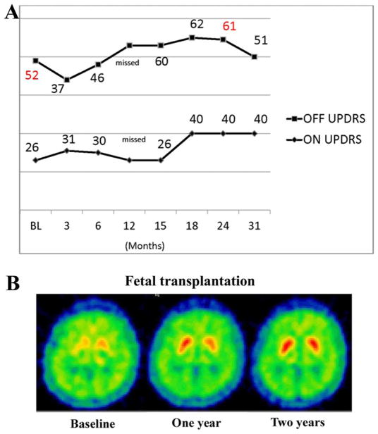
Methods: A 55-year-old levodopa-responsive woman with PD received bilateral putaminal fetal mesencephalic grafts as part of an NIH-sponsored double-blind sham-controlled trial. The patient never experienced clinical benefit, and her course was complicated by the development of graft-related dyskinesias. Fluorodopa positron emission tomography demonstrated significant increases postgrafting bilaterally. She experienced worsening of parkinsonism with severe dyskinesias, and underwent subthalamic nucleus deep brain stimulation 8 years after grafting. She died 16 years after transplantation.

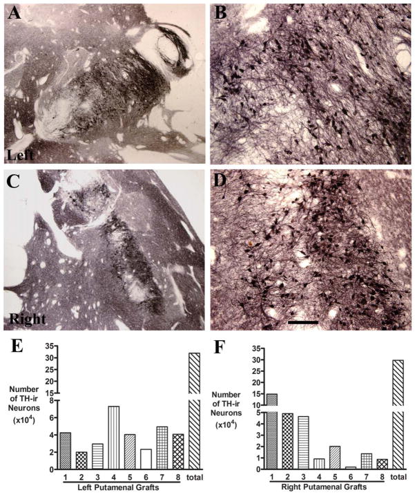
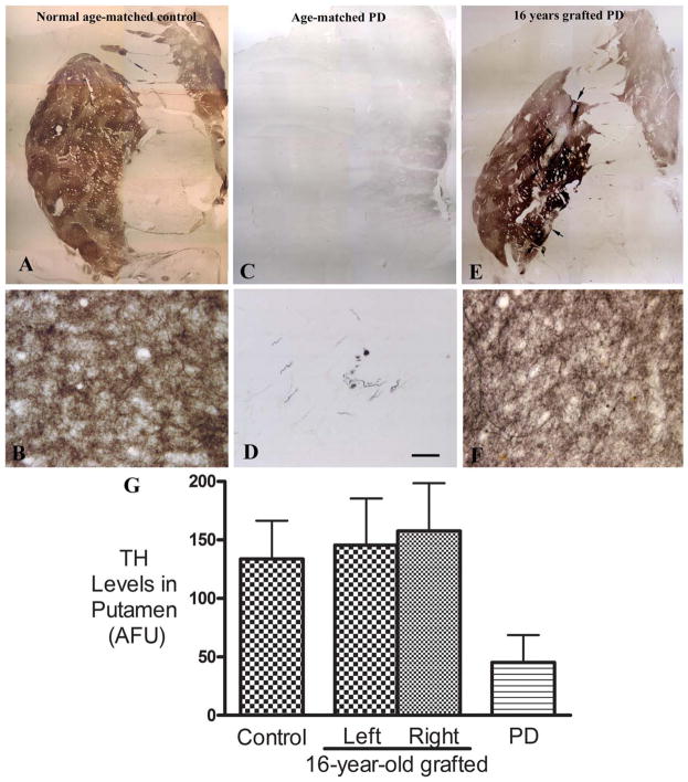
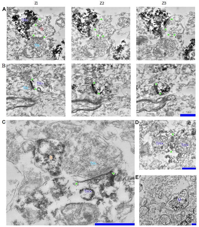
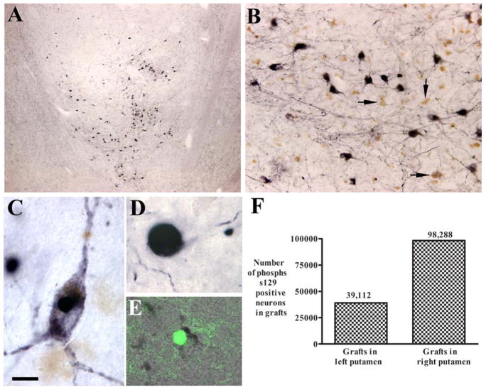
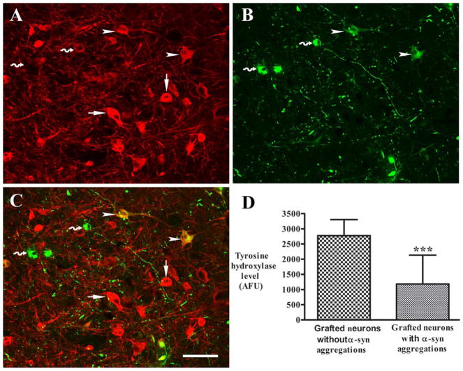
Results: Postmortem analyses confirmed the diagnosis of PD and demonstrated >300,000 tyrosine hydroxylase (TH)-positive grafted cells per side with normalized striatal TH-immunoreactive fiber innervation and bidirectional synaptic connectivity. Twenty-seven percent and 17% of grafted neurons were serine 129-phosphorylated α-synuclein positive in the left and right putamen, respectively.

Interpretation: These findings represent the largest number of surviving dopamine neurons and the densest and most widespread graft-mediated striatal dopamine reinnervation following a transplant procedure reported to date. Despite this, clinical recovery was not observed. Furthermore, the grafts were associated with a form of dyskinesias that resembled diphasic dyskinesia and persisted in the off-medication state. We hypothesize that the grafted cells produced a low level of dopamine sufficient to cause a levodopa-independent continuous form of diphasic dyskinesias, but insufficient to provide an antiparkinsonian benefit. ANN NEUROL 2017;81:46-57.


Conflict of interest statement

Potential Conflicts of Interest

Nothing to report.

Figures

FIGURE 1
FIGURE 1
(A) Blinded clinical assessment of motor function using Unified Parkinson Disease Rating Scale (UPDRS) part III in OFF and ON state at baseline and over 31 months postgrafting. (B) Positron emission tomography images at baseline, and 1 and 2 years postgrafting showing enhanced fluorodopa uptake and binding following dopaminergic transplantation.
FIGURE 2
FIGURE 2
(A–D) Low-power (A, C) and high-power (B, D) photomicrographs of putamen showing the robust survival of tyrosine hydroxylase–immunoreactive (TH-ir) grafted neurons in the right and left hemisphere. Bar in D=0.5mm in A–D. (E, F) Histograms showing the number of TH-ir neurons in individual grafts as well as the total number.
FIGURE 3
FIGURE 3
(E–F) Comparison of tyrosine hydroxylase (TH) immunoreactivity in putamen from a normal age-matched control without Parkinson disease (PD; A, B), an age- and Hoehn and Yahr score (HY)-matched PD patient (C, D; from the Rush PD Brain Repository), and our PD patient, 16 years after fetal cell grafting (E, F). The grafted putamen displayed extensive and intense TH immunoreactivity similar in intensity, volume, and pattern to the age-matched control. At higher magnification, dense fine TH-immunoreactive (TH-ir) fibers consisted of the fine network in gray matter of putamen (F). In the nongrafted age- and HY-matched PD brain, TH-ir fibers were not visible throughout much of the putamen. Bar in D=24 μm in B, D, and F, 2.85mm in A, C, and E. (G) Histogram showing the relative levels (mean+standard error of the mean [SEM]) of TH-ir fibers in putamen from the 3 brains shown in Figure 3A–F: an age-matched non-PD control, an age- and HY-matched PD case, and our PD patient 16 years after fetal cell grafting. SEM represents variability from different sections. AFU=arbitrary fluorescence units.
FIGURE 4
FIGURE 4
(A, B) (2) Serial section electron micrographs depicting (1) a host, tyrosine hydroxylase–negative (TH−) dendrite postsynaptic to a graft, TH-positive (TH+), bouton as well as (3) a host, TH−, bouton (A); TH+, spine synapsing with a host, TH+, bouton (B). (C) Host bouton synapsing onto a grafted dendritic shaft, positioned next to a dendritic spine (S). (D) Two TH+ grafted neurons synapsing onto each other. (E) TH+ myelinated axon (M) in close proximity to several host myelinated axons. Postsynaptic densities are denoted with arrowheads. Scale bars=0.5 μm for A, B, D, and E; 0.2 μm for C.
FIGURE 5
FIGURE 5
(A–E) Low-power (A) and high-power (B–E) microphotographs illustrating serine 129 phosphorylated α-synuclein–immunoreactive (S129-αSyn-ir; A–D) and thioflavin staining (E) pattern in the graft. Grafted neurons displayed S129-αSyn immunoreactivity (A, B). Some neurons exhibited dark soma and processes (B, C), and others were round (B, D). Many grafted neurons displayed neuromelanin without S129-αSyn immunoreactivity (B, arrows). Some grafted neurons displayed thioflavin aggregation (E). (F) In the grafted neurons, 27.11 % (right) and 10.45% (left) were S129-αSyn-ir. Bar in C=12 μm in C and E, 260 μm in A, 60μm in B, and 6 μm in D.
FIGURE 6
FIGURE 6
(A–C) Confocal microscopic images of a graft showing immunostaining pattern of tyrosine hydroxylase (TH; A, red) and serine 129 phosphorylated α-synuclein (S129-αSyn; B, green), and colocalization of TH and S129-αSyn (C, merged). Neurons with S129-αSyn inclusions had undetectable TH (curved arrows), and neurons with nonaggregated monomeric S129-αSyn (arrowheads) had diminished TH intensity compared with neurons without S129-αSyn immunoreactivity (arrows). (D) Quantification of relative fluorescent intensities revealed that optical densities of TH were significantly reduced in neurons with S129-αSyn inclusions relative to neurons with absent of S129-αSyn immunoreactivity. *** denotes p < .001. Bar in C=110 μm in A–C. AFU=arbitrary fluorescence units.

References

    1. Stenevi U, Björklund A, Svendgaard NA. Transplantation of central and peripheral monoamine neurons to the adult rat brain: techniques and conditions for survival. Brain Res. 1976;114:1–20. - PubMed
    1. Perlow MJ, Freed WJ, Hoffer BJ, et al. Brain grafts reduce motor abnormalities produced by destruction of nigrostriatal dopamine system. Science. 1979;204:643–647. - PubMed
    1. Björklund A, Stenevi U. Reconstruction of the nigrostriatal dopamine pathway by intracerebral nigral transplants. Brain Res. 1979;177:555–560. - PubMed
    1. Freed WJ, Perlow MJ, Karoum F, et al. Restoration of dopaminergic function by grafting of fetal rat substantia nigra to the caudate nucleus: long-term behavioral, biochemical, and histochemical studies. Ann Neurol. 1980;8:510–519. - PubMed
    1. Bakay RA, Barrow DL, Fiandaca MS, et al. Biochemical and behavioral correction of MPTP Parkinson-like syndrome by fetal cell transplantation. Ann N Y Acad Sci. 1987;495:623–640. - PubMed

Publication types