. 2016 Nov 30:7:13515.
doi: 10.1038/ncomms13515.

Structural decoding of netrin-4 reveals a regulatory function towards mature basement membranes

Affiliations

Structural decoding of netrin-4 reveals a regulatory function towards mature basement membranes

Raphael Reuten et al. Nat Commun. .

Abstract

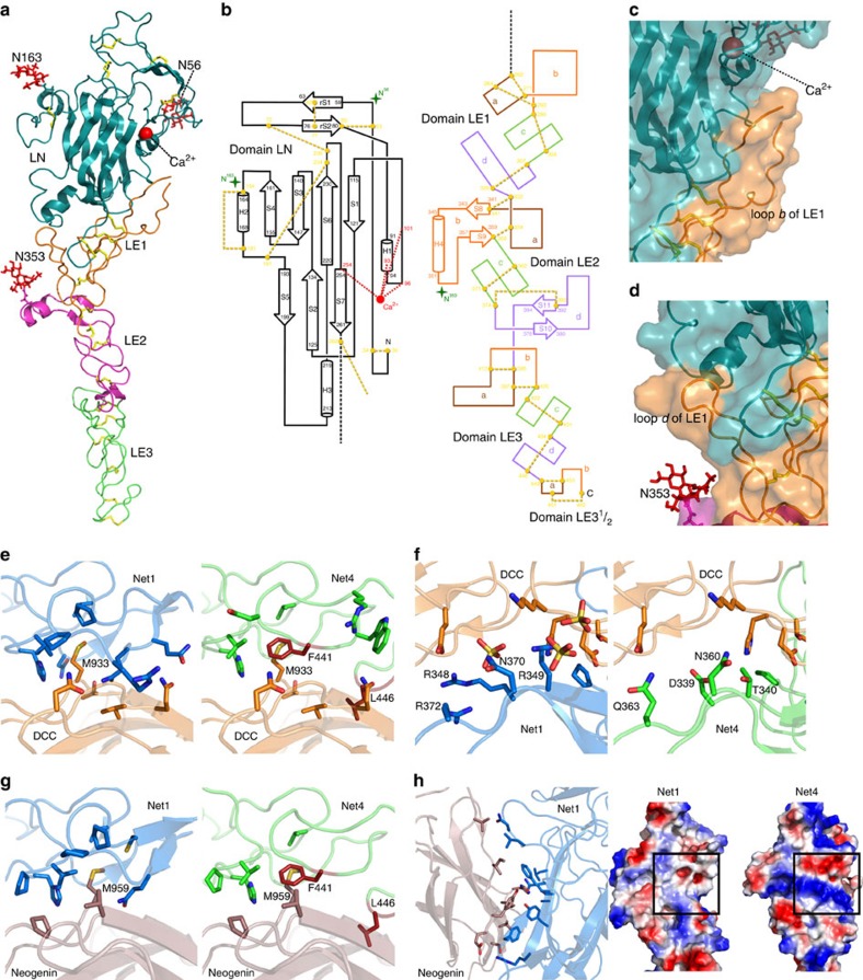
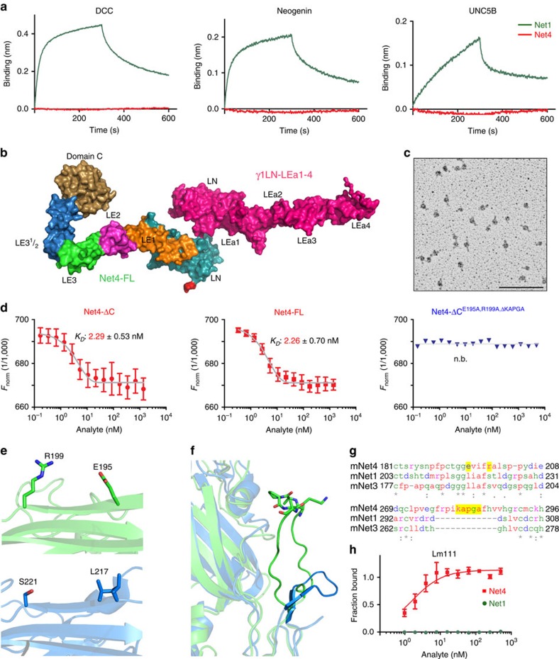
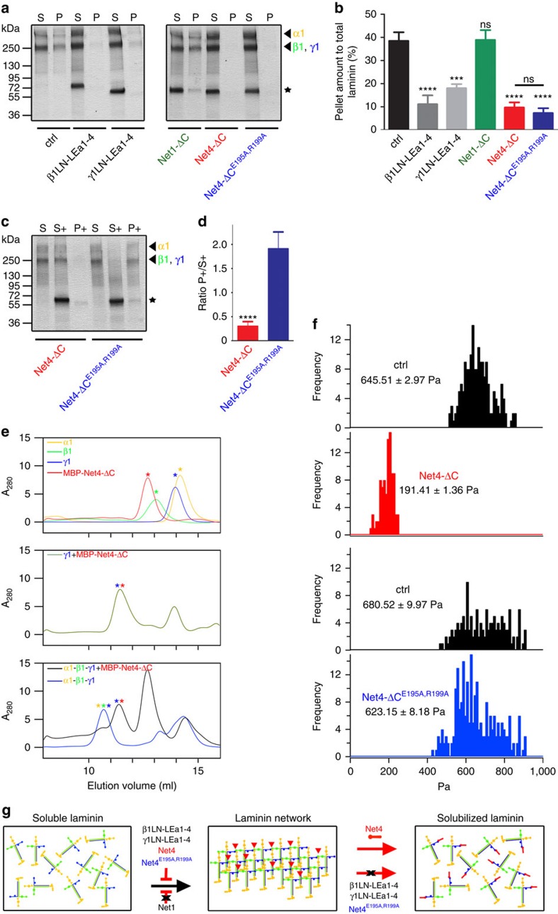
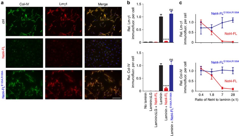
Netrins, a family of laminin-related molecules, have been proposed to act as guidance cues either during nervous system development or the establishment of the vascular system. This was clearly demonstrated for netrin-1 via its interaction with the receptors DCC and UNC5s. However, mainly based on shared homologies with netrin-1, netrin-4 was also proposed to play a role in neuronal outgrowth and developmental/pathological angiogenesis via interactions with netrin-1 receptors. Here, we present the high-resolution structure of netrin-4, which shows unique features in comparison with netrin-1, and show that it does not bind directly to any of the known netrin-1 receptors. We show that netrin-4 disrupts laminin networks and basement membranes (BMs) through high-affinity binding to the laminin γ1 chain. We hypothesize that this laminin-related function is essential for the previously described effects on axon growth promotion and angiogenesis. Our study unveils netrin-4 as a non-enzymatic extracellular matrix protein actively disrupting pre-existing BMs.


Figures

Figure 1
Figure 1. Overall structure of Net4.
(a) Ribbon model of Net4-ΔC viewed in the ‘open face' orientation. The domains LN (teal), LE1 (orange) LE2 (magenta) and LE3-31/2 (green) are coloured accordingly. N-linked glycans are drawn as red sticks, disulfide bridges are drawn as yellow sticks, and the calcium ion as a red sphere. (b) Secondary structure diagrams. Disulfide connectivity in domains LN and LE are indicated by yellow dotted lines, visible N-linked glycans by green stars with the Asn residues N56, N163 and N353 highlighted. β-strands are depicted as arrows and the α-helices as cylinders, each labelled with the first and last residue. The individual loop segments are shown in different colour codes. (c) Interface between domains LE1 and LN. The KAPG motif is free in solution and not visible in the structure. (d) Secondary interface between domains LE1 and LN. The loop d segment between Cys307 and Cys329 of subdomain LE1 is in contact with the N-terminus and the loop S5-H3 of domain LN. (e) Complex of Net1 (marine; PDB 4OVE) and DCC (orange; PDB 4URT) at site 1 with interacting residues shown at full opacity and the same complex with Net4 (green) replacing Net1. (f) Complex of Net1 (marine) and DCC (orange; PDB 4URT) at site 2 with interacting residues shown at full opacity and the same complex with Net4 (green) replacing Net. (g) Complex of Net1 (marine) and neogenin (pink; PDB 4PLN) at site 1 with interacting residues shown at full opacity and the same complex with Net4 (green) replacing Net1. Similar to site 1 of the DCC-Net1 complex, F441 (red) of Net4 occludes Met959 of neogenin from the hydrophobic pocket. L456 (red) of Net4 again clashes with the binding partner. (h) Net1 (marine) in complex with neogenin (pink) at site 2. Note the difference in the surface charge at the neogenin binding site (indicated by a box) of Net1 (left) and the corresponding site on Net4 (right).
Figure 2
Figure 2. Binding comparison of Net4 and Net1 to netrin receptors and laminin.
(a) Biolayer interferometry binding kinetic analysis of DCC, neogenin, and UNC5B to Net4 (red) as well as Net1 (green). AHC sensors were coated with the Fc chimera proteins. Association (from 0 to 300 s) and dissociation (from 300 to 600 s) phases are shown for 50 nM of both netrins. (b) The SAXS model for the Net4-FL-γ1LN-LEa1-4 complex was generated by firstly calculating models for Net4-ΔC-γ1LN-LEa1-4 complex (Supplementary Fig. 5d) followed by the addition of the LE and globular domain C. This model highlights the role of N-terminal globular domains in the interaction. The agreement between experimentally collected SAXS data and model-derived SAXS data is presented in Supplementary Table 1. (c) Rotary-shadowing electron microscopy image of Net4-ΔC-γ1LN-LEa1-4 clearly showing a 1:1 complex mediated via the globular domains. (d) Microscale thermophoresis binding analysis of different Net4 mutants to labelled γ1LN-LEa1-4. Binding of Net4 lacking the netrin-specific C domain (Net4-ΔC). Binding of full-length Net4 (Net4-FL). Analysis of the binding of a Net4 mutant (Net4-ΔCΔKAPGA) in which the specific loop b (KAPGA) within LE1 was deleted. Binding of a combined Net4 mutant (Net4-ΔCE195A,R199A,ΔKAPGA). Error bars, s.d. (n=3 independent technical replicates). KD values are shown in the graph (n.b., no binding). (e,f) Laminin-binding epitopes within Net4 (green) compared with the equivalent positions in Net1 (marine). Note loop b of LE1 of Net4 has an extensive contact area with the LN domain, and the KAPGA motif (shown in stick notation) is highly flexible. (g) Sequence alignment of Net4, Net1 and netrin-3 showing the laminin-binding region. (h) Solid-phase binding study of Net4 and Net1 to immobilized laminin γ1 (γ1LN-LEa1-4). Error bars, s.d. (n=3 independent technical replicates). Scale bar, (c) 100 nm.
Figure 3
Figure 3. Net4 disrupts pre-existing laminin networks.
(a) Inhibition of laminin 111 polymerization (Lm111) by human laminin β1 (β1LN-LEa1-4), mouse laminin γ1 (γ1LN-LEa1-4), mouse Net1 (Net1-ΔC), mouse Net4-ΔC and the laminin-binding mutant mouse Net4-ΔCE195A,R199A. For the experimental scheme depicting the laminin 111 polymerization assay see Supplementary Fig. 7a. SDS–PAGE analysis of the pellet (P) and the supernatant fraction (S). The position of the laminin chains (arrow heads) and the recombinant proteins (stars) are indicated. (b) Densitometric analysis of three independent polymerization experiments and the percentage of the pellet fraction are displayed. (mean±s.d.; n=3; ****P<0.00006 (ctrl vs β1LN-LEa1-4, Net4-ΔC and Net4-ΔCE195A,R199A) and ***P=0.0004 (ctrl vs γ1LN-LEa1-4); ns, not significant). (c) For the method procedure see Supplementary Fig. 7b. The resulting fractions (supernatant of laminin polymerization (S), supernatant after addition of different proteins (S+) and polymer (P+)) were separated by SDS–PAGE, followed by Coomassie Brilliant-Blue staining. Different laminin chains are colour labelled (α1 (yellow), β1 (green) and γ1 (blue)) and added recombinant proteins are indicated with asterisks. (d) The ratio between polymer (P+) and supernatant (S+) was determined by densitometric analysis of at least three independent experiments (mean±s.d.; n=3; ****P=0.00005). (e) N-terminal laminin fragments (LN-LEa1-4) of α1 (yellow), β1 (green) and γ1 (blue) as well as MBP-Net4-ΔC (red) single proteins were analysed using analytical size-exclusion chromatography (top). SEC profile of the laminin fragment γ1LN-LEa1-4 (γ1) in complex with MBP-Net4-ΔC (green, middle). Complexes [α1-β1-γ1 (blue) and α1-β1-γ1+MBP-Net4-ΔC (red)] were analysed by SEC (bottom) revealing that the MBP-Net4-ΔC is able to disrupt the α1-β1-γ1 complex. Asterisks indicate the single proteins within the complex. (f) Atomic force microscopy anaylsis of a polymerized Matrigel matrix treated with Net4-ΔC or with the laminin-binding mutant Net4-ΔCE195A,R199A. (g) Model depicting inhibition of laminin polymerization via laminin β1 (β1LN-LEa1-4), laminin γ1 (γ1LN-LEa1-4), mouse Net4-ΔC, and the laminin-binding mutant mouse Net4-ΔCE195A,R199A but not via mouse Net1 (Net1-ΔC). The pre-existing laminin network can only be solubilized through Net4. Error bars, s.d. (n=3 independent technical replicates).
Figure 4
Figure 4. Net4 blocks laminin assembly on the cell surface.
(a) Within untreated (ctrl), Net4-FL-, and Net4-FLE195A,R199A-treated (28-fold excess in molarity compared with laminin) Schwann cell extracellular matrix assembly assay BM components collagen IV (Col-IV, green) and laminin 111 (Lmγ1, red) were stained. Cells were counter-stained for DAPI (blue). (b) Staining intensities are displayed relative to the DAPI signal [relative Lmγ1 intensity per cell (left) and relative Col-IV intensity per cell, (right)] (mean±s.d.; n=5; ****P<0.00001; ns, not significant). Error bars, s.d. (n=5 independent cell cultures). (c) Concentration dependent effect of Net4-FL and Net4-FLE195A,R199A on BM formation indicated through measurement of the relative Lmγ1 intensity/cell (left) and relative Col-IV intensity/cell (right). Net4 proteins were added together with the BM components laminin 111, collagen IV and nidogen-1 with increasing molar ratios to laminin 111 (0.4, 1.8, 7, 28-fold excess). Scale bars, (a) 200 μm. P values were calculated by one-way ANOVA followed by a pairwise comparison using the Holm-Sidak method. ANOVA, analysis of variance.
Figure 5
Figure 5. Net4 induces neurite outgrowth in a laminin-dependent manner.
(a) Diagram depicting the olfactory bulb (OB) explants assay procedure. (b) Representative images of untreated (ctrl), laminin γ1 (γ1LN-LEa1-4)-, Net4 (Net4-ΔC)-, Net4 in combination with laminin γ1 (Net4-ΔC+γ1LN-LEa1-4)-, and Net4 laminin-binding mutant (Net4-ΔCE195A,R199A)-treated OB explants. (c) Statistical analysis of OB explants (mean±s.e.m.; n=19; ****P<0.0001, ns, not significant). Error bars, s.e.m. (n=19 independent cell cultures). P values, two sided t-test. Scale bar, 200 μm (b).
Figure 6
Figure 6. Net4 activity on angiogenesis is dependent on Net4/laminin interaction.
(a) Analyses of tube-like structure inhibition by Net4-ΔC mutants (Net4-ΔCE195A, Net4-ΔCR199A and Net4-ΔCΔKAPGA) impaired in laminin γ1 binding (protein concentrations: 1 μM). Statistical analyses of tube formation via determining the number of cell cluster connections (tube number) (mean±s.d.; n=3; ***P=0.0007) (right). Error bars, s.d. (n=3 independent cell cultures). (b) Blocking of Net4-ΔC inhibited tube formation through equimolar addition of γ1LN-LEa1-4 (1 μM, molar ratio of Net4-ΔC:γ1LN-LEa1-4, 1:1). The laminin fragment γ1LN-LEa1-4 competes with Net4 binding to laminin (mean±s.d.; n=3; *P=0.035; ns, not significant). Error bars, s.d. (n=3 independent cell cultures). (c) Treatment of VEGF-A induced spheroid sprouting of HDMECs embedded in collagen I by Net4-ΔC (1 μM). Analysis of sprouting events (ns, not significant). (d) Tube formation analyses of co-cultured endothelial and perivascular-like cells from untreated and Net4-FL- or Net4-FLE195A,R199A-treated cultures (30 nM). Representative images of CD31 staining are shown. The tube length (mm per mm2) was quantified through the CD31 staining (mean±s.d.; n=5; ****P<0.00005). (e) Apotome images of Col-IV, Lmγ1 and CD31 stained tubes from control (ctrl), Net4-FL and Net4-FLE195A,R199A treatment are displayed. Error bars, s.d. (n=3, (ac); n=5, (d,e) independent cell cultures). P values, two sided t-test. Scale bars, 100 μm (ad); 50 μm (e).
Figure 7
Figure 7. Laminin polymers are essential to maintain capillary networks and tumour progression.
(a) Visualization of capillaries via FITC-dextran injection of untreated (ctrl), Net4-FL- and Net4-FLE195A,R199A-treated CAMs after 48 h. The vascularized area from different protein treatments (left) was determined (see Methods section) at 48 h (mean±s.d.; n=5; ****P<0.0001). Error bars, s.d. (n=5 independent cell cultures). (b) H&E staining of untreated (ctrl) and Net4-FL-, and Net4-FLE195A,R199A-treated CAMs after 48 h. Red lines and red arrow heads indicate the capillary density (capillaries are labelled with C). (c) Diagram showing the tumour treatment regimen. (d) Macroscopic images of melanoma treated daily with 1 μM of control protein (mouse serum albumin, left), Net4-FL (middle), Net4-FLE195A,R199A (right). (e) Progression of melanoma treated with Net4-FL and the laminin γ1 binding mutant Net4-FLE195A,R199A (mean±s.d.; n=5; ****P<0.0001). Error bars, s.d. (n=5 animals, female C57BL/6). P values, two sided t-test. Scale bars, 100 μm (a); 20 μm (b); and 5 mm (d).
Figure 8
Figure 8. Impact of Net4 on disruption of the laminin network and its effect on vascularization.
(a) Diagram indicates vessel diameter distribution (left) within Net4-FLE195A,R199A-treated CAMs (blue squares), and the Net4-FL group (red triangle). Model showing the common established capillary hierarchy formed within the CAM. Numbers indicate sizes of different vessels in the common established hierarchy (middle). The table displays the diameter of different hierarchical vessels from Net4-FLE195A,R199A- (blue) and Net4-FL-treated (red) experiments (right) (n.p., not present). (b) Ultrastructural analyses of Net4-FLE195A,R199A- (blue) and Net4-FL-treated (red) CAMs using transmission electron microscopy. Images show the capillary endothelium surrounded by perivascular cells. The endothelium E is shown and PVCs are indicated as P. Electron microscopy images highlight the vascular basement membrane (vBM, red arrow heads (middle) and red asterisks (bottom)) between the endothelium and PVCs, which are surrounded by a vBM layer. (c) Model of Net4 effect on capillary cell types (endothelium (Endoth.) and pericyte (Pericyte)) and the vBM. Scale bars (b), 2 μm (top); 1 μm (middle); 0.5 μm (bottom).

Similar articles

Cited by

References

    1. Ishii N., Wadsworth W. G., Stern B. D., Culotti J. G. & Hedgecock E. M. UNC-6, a laminin-related protein, guides cell and pioneer axon migrations in C. elegans. Neuron 9, 873–881 (1992). - PubMed
    1. Kennedy T. E., Serafini T., de la Torre J. R. & Tessier-Lavigne M. Netrins are diffusible chemotropic factors for commissural axons in the embryonic spinal cord. Cell 78, 425–435 (1994). - PubMed
    1. Koch M. et al.. A novel member of the netrin family, beta-netrin, shares homology with the beta chain of laminin: identification, expression, and functional characterization. J. Cell Biol. 151, 221–234 (2000). - PMC - PubMed
    1. Nakashiba T. et al.. Netrin-G1: a novel glycosyl phosphatidylinositol-linked mammalian netrin that is functionally divergent from classical netrins. J Neurosci 20, 6540–6550 (2000). - PMC - PubMed
    1. Nakashiba T., Nishimura S., Ikeda T. & Itohara S. Complementary expression and neurite outgrowth activity of netrin-G subfamily members. Mech. Dev. 111, 47–60 (2002). - PubMed

Publication types

MeSH terms