. 2016 Nov 30:6:37990.
doi: 10.1038/srep37990.

Structural basis for the dissociation of α-synuclein fibrils triggered by pressure perturbation of the hydrophobic core

Affiliations

Structural basis for the dissociation of α-synuclein fibrils triggered by pressure perturbation of the hydrophobic core

Guilherme A P de Oliveira et al. Sci Rep. .

Abstract

Parkinson's disease is a neurological disease in which aggregated forms of the α-synuclein (α-syn) protein are found. We used high hydrostatic pressure (HHP) coupled with NMR spectroscopy to study the dissociation of α-syn fibril into monomers and evaluate their structural and dynamic properties. Different dynamic properties in the non-amyloid-β component (NAC), which constitutes the Greek-key hydrophobic core, and in the acidic C-terminal region of the protein were identified by HHP NMR spectroscopy. In addition, solid-state NMR revealed subtle differences in the HHP-disturbed fibril core, providing clues to how these species contribute to seeding α-syn aggregation. These findings show how pressure can populate so far undetected α-syn species, and they lay out a roadmap for fibril dissociation via pathways not previously observed using other approaches. Pressure perturbs the cavity-prone hydrophobic core of the fibrils by pushing water inward, thereby inducing the dissociation into monomers. Our study offers the molecular details of how hydrophobic interaction and the formation of water-excluded cavities jointly contribute to the assembly and stabilization of the fibrils. Understanding the molecular forces behind the formation of pathogenic fibrils uncovered by pressure perturbation will aid in the development of new therapeutics against Parkinson's disease.


Figures

Figure 1
Figure 1. Initial characterization of α-syn monomers and fibrils for the HHP-NMR studies.
(a) 1H-15N HSQC spectra of α-syn (αS) monomers. (b) The washing/centrifugation (w/c) steps were indirectly probed by dot-blots to exclude any remaining oligomeric species formed during the fibrillation reaction. We used eight w/c steps to decrease the amount of oligomers remaining in the fibril samples, as determined by the A-11 antibody signal. (c) Quantification of dot-blots was performed by densitometric analysis using the ImageJ software. The results are shown as the avg ± s.d. of three independent experiments.
Figure 2
Figure 2. Species released from α-syn fibrils under HHP.
(ac) HHP-disturbed fibrils at 1 bar. (a) SEC in Superdex 200 10/300 of fibrils after 1 h of treatment at 516, 1,033, and 2,067 bar. Absorbance at 214 nm of monomeric α-syn peak increased as HHP increments were applied to fibrils. (b) ThT fluorescence signal of fibrils (αS-F) decreased similarly to the signals of monomers (αS-M) after 1 h at 1,033 bar. (c) The ellipticity of fibrils subjected to HHP shifted from β-shift secondary structure (negative peak at 220 nm) to a random conformation (negative peak at 200 nm), which is typical of α-syn monomers. (d) Disassembly kinetics of sedimented fibrils at 1,033 (black) and 2,067 (red), as monitored by the loss of ThT fluorescence. Sedimented fibrils were subjected to these pressure values for the whole kinetics immediately after ThT signal stabilization at 1 bar, which was achieved after 3 h. Measurements are shown as avg. ± s.e.m. (n = 3, independent protein batches). (e) Visual inspection of ThT signal under UV light of non-sedimented fibrils (before) and sedimented fibrils (after 3 h at 1 bar) and after ca. 6 h subjected to 516, 1,033, 2,067, and 3,100 bar of pressure increments. (f) The absorbance of the amide I′ band as a function of pressure increments of15N/13C α-syn fibrils by HHP-FTIR. The pressure ranges from 1 to 3,121 bar and from 3,538 to 7,228 bar are shown in black and red, respectively, to highlight the shifted peaks. (g) Amide I′ band recovery at 37 °C was monitored by FTIR spectroscopy every 30 min after pressure release for 360 min. Red and gray lines represent the shift recovery over time. The dashed gray line representsthe amide I’ band after 12 h. (h) Ellipticity response over time to 516 bar, as measured by HHP-CD.
Figure 3
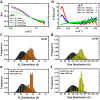
Figure 3. Small-angle X-ray scattering profile of α-syn species released from fibrils.
(a) Scattering intensity I(s) as a function of the scattering vector s of α-syn fibrils (αS-F) at 1 and after 516, 1,033, and 2,067 bar for 1 h. Scattering was measured at atmospheric pressure. (b) Kratky plots showing the shift of the folding pattern of fibrils to the flexible and unfolded behavior of species dissociated from fibril safter HHP treatment. (cf) Frequency of radius of gyration, Rg (orange), and size distribution (yellow) of initial monomers (c,d) and species released from fibrils after 1 h of 2,067 bar treatment (e,f), as evaluated by the ensemble optimization method. Black bars represent the distribution of the pool of conformers (a collection of 10,000 random conformers) used to define the conformational space.
Figure 4
Figure 4. α-Syn fibril dissociation into monomers, as monitored by HHP-NMR.
(a) Superposition of 1H-15N HSQC spectra of monomeric α-syn (αS-M) at different pressure increments ranging from 1 to 2,500 bar. The systematic chemical shifts of the1H-15N correlations with increasing pressure are the result of the compressibility effects pressure imposes on the solvent hydrogen bonds to the protein backbone. (b) HHP titration of α-syn fibrils (αS-F) using pressure increments higher than 500 bar revealed 1H-15N correlations typical of the monomeric protein. (c) Line-broadening analysis of the response to increasing pressure for monomeric α-syn (black lines) and for monomeric species released from the fibril (blue lines). Each line corresponds to one 1H-15N correlation in which we were able to follow the line-broadening behavior as a function of increasing pressure. (d) The r-square (R2) values and the second-order coefficients (b2) as a function of residues obtained after fitting the bell-shaped behavior of the α-syn species released from fibrils to a second-order polynomial equation.
Figure 5
Figure 5. Conformational dynamics of monomeric species released from HHP-disturbed fibrils.
Relaxation rates (R2 eff) obtained from the intensities of resonance in 1H-15N correlation spectra acquired with 50 (open symbols) and 1000 Hz (filled symbols) for initial monomers (αS-M, blue) at 1, 500 and 750 bar and from species released from fibrils (αS from αS-F, red) at the same pressure values. All experiments with monomeric α-syn and fibrillar samples were taken at the specified pressure after 20 minutes of equilibration. Experiments were run once and error bars represent deviations from noise peaks.
Figure 6
Figure 6. HHP effects on fibril structure.
(a) Solid-state NMR structure of full-length α-syn monomer (PDB: 2n0a) highlighting the N- and C-terminus (blue) and the Greek-key arrangement of the core (green). (b) Solid-state NMR structure of the α-syn fibril core (residues 46–96, PDB: 2n0a) showing the key elements for fibril stability: the salt-bridge between E46-K80 (gray), steric zippers involving V49, V77 and V82 (cyan), the glutamine ladder Q79 (purple) and the hydrophobic packing involving I88, A91 and F94 (orange). Threonine positions (yellow) are highlighted to show the effects of HHP on the α-syn fibril core. (c) Negatively stained transmission electron microscopy of fibrils before and after 1 h treatment at 1,033 bar. (d) 13C–13C correlation spectrum of the α-syn fibril core acquired in a static magnetic field of 600 MHz before (blue) and after (red) 1 h at 1,033 bar. Dashed lines show the changes.
Figure 7
Figure 7. Seeding of remaining fibrils after HHP treatment.
Aggregation kinetics of α-syn monomers (αS-M) in the absence of seeds (black spheres) or in the presence of increasing micrograms of (a) sonicated fibrils and (b) structural modified monomers (SMMs) + remaining fibrils formed after 1 h incubation of fibrils at 1,033 bar. Measurements of the ThT fluorescence are shown as avg. ± s.d. of three independent experiments. Insets show negatively stained electron microscopy images of seeds used for the kinetic experiments.
Figure 8
Figure 8. Mechanism of dissociation of α-syn fibrils by pressure.
(a,b) The cavity-prone hydrophobic core of α-syn fibrils (PDB: 2n0a). Non-exposed cavities along the fibril axis and residues located in the vicinity of these cavities are highlighted as green surfaces and gray sticks, respectively. (c) Hydration of the hydrophobic core triggered by pressure. The cavity that may sense hydration first (purple) is the one formed by the side chains of the E46-K80 pair, followed by the ones located inward toward the cavity-prone hydrophobic bulk (orange). The presence of other small cavities (green) may ultimately disrupt the fibril core. (d) Light scattering (measured as the area under the light scattering curve A and normalized by the value at 1 bar A0) of α-syn fibrils in the absence or in the presence of 20 and 30% of glycerol. Measurements are shown as avg. ± s.e.m. (n = 3, independent protein preparations). (e) Schematic representation of pressure effects on α-syn fibrils, showing the major findings obtained by challenging α-syn fibrils with pressure. Structurally modified monomers (SMMs) are shown in red and remaining fibrils in blue.

Similar articles

Cited by

References

    1. Spillantini M. G. et al.. Alpha-synuclein in Lewy bodies. Nature 388, 839–840 (1997). - PubMed
    1. Polymeropoulos M. H. et al.. Mutation in the alpha-synuclein gene identified in families with Parkinson’s disease. Science 276, 2045–2047 (1997). - PubMed
    1. Krüger R. et al.. Ala30Pro mutation in the gene encoding alpha-synuclein in Parkinson’s disease. Nat. Genet. 18, 106–108 (1998). - PubMed
    1. Singleton A. B. et al.. alpha-Synuclein locus triplication causes Parkinson’s disease. Science 302, 841 (2003). - PubMed
    1. Chartier-Harlin M. C. et al.. Alpha-synuclein locus duplication as a cause of familial Parkinson’s disease. Lancet 364, 1167–1169 (2004). - PubMed

Publication types

MeSH terms

LinkOut - more resources