. 2017 Jan 3;8(1):1092-1109.
doi: 10.18632/oncotarget.13582.

Spermidine/spermine N1-acetyltransferase regulates cell growth and metastasis via AKT/β-catenin signaling pathways in hepatocellular and colorectal carcinoma cells

Affiliations

Spermidine/spermine N1-acetyltransferase regulates cell growth and metastasis via AKT/β-catenin signaling pathways in hepatocellular and colorectal carcinoma cells

Cong Wang et al. Oncotarget. .

Abstract

Hepatocellular carcinoma (HCC) and colorectal cancer (CRC) are among the most common cancers across the world. Therefore, identifying the potential molecular mechanisms that promote HCC and CRC progression and metastasis are urgently needed. Spermidine/spermine N1-acetyltransferase (SSAT) is a catabolic enzyme that acetylates the high-order polyamines spermine and spermidine, thus decreasing the cellular content of polyamines. Several publications have suggested that depletion of intracellular polyamines inhibited tumor progression and metastasis in various cancer cells. However, whether and how SSAT regulates cell growth, migration and invasion in hepatocellular and colorectal carcinoma cells remains unclear. In this study, depletion of polyamines mediated by SSAT not only attenuated the tumor cell proliferation but also dramatically inhibited cell migration and invasion in hepatocellular and colorectal carcinoma cells. Subsequent investigations revealed introduction of SSAT into HepG2, SMMC7721 hepatocellular carcinoma cells and HCT116 colorectal carcinoma cells significantly suppressed p-AKT, p-GSK3β expression as well as β-catenin nuclear translocation, while inhibition of GSK3β activity or exogenous polyamines could restore SSAT-induced decreases in the protein expression of p-AKT, p-GSK3β and β-catenin. Conversely, knockdown of SSAT in Bel7402 hepatocellular carcinoma cells and HT-29 colorectal carcinoma cells which expressed high levels of SSAT endogenously significantly promoted the expression of p-AKT, p-GSK3β as well as β-catenin nuclear translocation. Taken together, our results indicated depletion of polyamines by SSAT significantly inhibited cell proliferation, migration and invasion through AKT/GSK3β/β-catenin signaling pathway in hepatocellular carcinoma and colorectal cancer cells.

Keywords: AKT; SSAT; metastasis; polyamine; β-catenin.


Conflict of interest statement

CONFLICTS OF INTEREST

The authors declare that they have no competing interest.

Figures

Figure 1
Figure 1. SSAT over-expression down-regulated the content of polyamines in hepatocellular and colorectal carcinoma cells
A. Western blot analysis determined the level of SSAT in three hepatocellular carcinoma cell lines (Hep G2, SMMC7721 and Bel7402) and two colorectal carcinoma cell lines (HCT 116, HT-29). B. Western blot analysis demonstrated that SSAT was upregulated in Hep G2, SMMC7721 and HCT 116 cells after stably transfection. Two stably transfected cell clonies were selected in each cell lines. β-actin was used as internal control. C. HPLC analysis showed the concentration of polyamines including spermidine and spermine were significantly down-regulated in HepG2, SMMC7721 and HCT116 cells transfected with SSAT compared with the cells transfected with Vector. While the acetylated polyamine including N1-acetylspermidine, N1-acetylspermine were profoundly increased in HepG2, SMMC7721 and HCT 116 cells transfected with SSAT compared with the cells transfected with Vector. All of the experiments were repeated three times and data was presented as means ± SEM (n=3). *p< 0.05, **p<0.01, ***p<0.001 compared to the vector group. #p< 0.05, ##p<0.01, ###p<0.001 compared to the vector group, one way ANOVA.
Figure 2
Figure 2. Knockdown of SSAT up-regulated the content of polyamines in hepatocellular and colorectal carcinoma cells
A. Western blot analysis demonstrated that SSAT was downregulated in Bel7402 and HT29 cells after treatment with two different specific SSAT siRNA. B. HPLC analysis showed spermidine and spermine were significantly up-regulated in Bel7402 and HT29 cells transfected with SSAT siRNA compared with the cells transfected with non-sense siRNA. While the acetylated polyamine including N1-acetylspermidine, N1-acetylspermine were profoundly decreased in Bel7402 and HT29 cells transfected with SSAT siRNA compared with the cells transfected with non-sense siRNA. All of the experiments were repeated three times and data was presented as means ± SEM (n=3). *p< 0.05, **p<0.01, ***p<0.001 compared to the vector group. #p< 0.05, ##p<0.01, ###p<0.001 compared to the vector group, one way ANOVA.
Figure 3
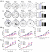
Figure 3. SSAT was involved in cell colony formation and proliferation in hepatocellular and colorectal carcinoma cells
A. Cell colony formation results showed enforced expression of SSAT inhibited the colony formation in HepG2, SMMC7721 and HCT 116 cells compared with cells transfected with Vector. B, C. MTT assay showed SSAT over-expression attenuated the cell proliferation in HepG2, SMMC7721 and HCT 116 cells, whereas knockdown of SSAT by siRNA enhanced cell proliferation in Bel7402 and HT-29 cells. Representative images were shown. All of the experiments were repeated three times and data was presented as means ± SEM (n=3). *, p< 0.05, **, p<0.01 and ***, p<0.001 compared to the control group.
Figure 4
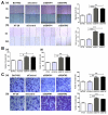
Figure 4. Up-regulation of SSAT inhibited cell migration and invasion in hepatocellular and colorectal carcinoma cells
A. Wound healing assay indicated that SSAT up-regulation promoted HepG2, SMMC7721 and HCT116 cells migration. B, C. Transwell chamber without (B) or with Matrigel (C) were employed to examine the migration and invasion ability of tumor cells. Cells were harvested and added in serum-free media into the top chambers. After 24 h migration or invasion, the cells on the lower surface of the filters were stained. Note that up-regulation of SSAT significantly enhanced cell migration and invasion in HepG2, SMMC7721 and HCT 116 cells. Representative images and the number of migrated or invaded cells were shown. All of the experiments were repeated at least three times and data was presented as means ± SEM. *p<0.05, **p<0.01, ***p<0.001 compared to the control group, one-way ANOVA.
Figure 5
Figure 5. Knockdown of SSAT enhanced cell migration and invasion in hepatocellular and colorectal carcinoma cells
A. Wound healing assays revealed that knockdown of SSAT inhibited cell migration in Bel7402 and HT-29 cells. B, C. Knockdown of SSAT attenuated the migration and invasion in Bel7402 and HT-29 cells. Bel7402 or HT-29 cells without transfection or transfected with non-sense siRNA or SSAT specific siRNA were seeded into the transwell chamber coated without (B) or with Matrigel (C) and incubated in RPMI1640 containing 10% FBS for 24 h. Representative images were shown. All of the experiments were repeated at least three times and data was presented as means ± SEM. *p< 0.05, **p<0.01, ***p<0.001 compared to the control group, one-way ANOVA.
Figure 6
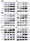
Figure 6. Up-regulation of SSAT inhibited AKT/GSK3β/β-catenin signaling pathway and Epithelial-to-Mesenchymal transition in hepatocellular and colorectal carcinoma cells
A. Western blot analysis demonstrated that the protein expression of p-AKT, AKT and p-GSK3 was decreased in HepG2, SMMC7721 and HCT 116 cells transfected with SSAT compared with the cells without transfection or transfected with Vector. β-actin was used as an internal control. B, C. Western blot analysis showed the expression of β-catenin (total and nucleus protein), cyclin D1, N-cadherin, Slug and Twist1 were decreased in SMMC7721, HepG2 and HCT 116 cells transfected with SSAT compared with the cells without transfection or transfected with Vector. The epithelial marker E-cadherin was increased in HepG2, SMMC7721 and HCT 116 cells transfected with SSAT compared with the cells without transfection or transfected with Vector. Lamin B1 was used as an internal control of nucleus protein, β-actin was used as an internal control of total protein.
Figure 7
Figure 7. Silencing of SSAT induced AKT/GSK3β/β-catenin signaling pathway and Epithelial-to-Mesenchymal transition in hepatocellular and colorectal carcinoma cells
A. Western blot analysis demonstrated that the protein expression of p-AKT, AKT and p-GSK3 was increased in Bel7402 and HT-29 cells transfected with SSAT siRNA compared with the cells transfected with non-sense siRNA. β-actin was used as an internal control. B, C. Western blot analysis showed the expression of β-catenin (total and nucleus protein), cyclin D1, N-cadherin, Slug and Twist1 was increased in Bel7402 and HT-29 cells transfected with SSAT siRNA compared with the cells transfected with non-sense siRNA. The expression of N-cadherin was decreased in Bel7402 and HT-29 cells transfected with SSAT siRNA. Lamin B1 was used as an internal control of nucleus protein, β-actin was used as an internal control of total protein.
Figure 8
Figure 8. The GSK3β inhibitor LiCl reversed SSAT-induced inhibition of AKT/β-catenin signaling pathway as well as cell proliferation, migration and invasion in hepatocellular and colorectal carcinoma cells
A. Western blot analysis showed GSK 3β inhibitor LiCl markedly reversed SSAT-induced p-GSK3β and β-catenin down-regulation in SMMC7721, HepG2 and HCT116 cells transfected with SSAT. β-actin served as an internal control. B. MTT assay showed LiCl attenuated SSAT-induced inhibition of cell proliferation in SMMC7721 (a), HepG2 (b) and HCT 116 cells (c). *p< 0.05, **p<0.01, ***p<0.001 compared to the control group, one-way ANOVA. C. Transwell assay with Matrigel demonstrated LiCl restored SSAT-induced inhibition of cell invasion in SMMC7721, HepG2 and HCT 116 cells. *p< 0.05, **p<0.01, ***p<0.001 compared to the control group, one-way ANOVA.
Figure 9
Figure 9. The exogenous polyamines (PUT, SPD, SPM) reversed SSAT-induced inhibition AKT/β-catenin signaling pathway as well as cell proliferation and invasion in hepatocellular and colorectal carcinoma cells
A. Western blot analysis showed exogenous polyamines reversed SSAT-induced p-AKT, AKT, p-GSK3β and β-catenin down-regulation in HepG2, SMMC7721 and HCT116 cells transfected with SSAT. β-actin served as an internal control. B. MTT assay showed exogenous polyamines attenuated SSAT-induced inhibition of cell proliferation in SMMC7721 (a), HepG2 (b) and HCT 116 cells (c). *p< 0.05, **p<0.01, ***p<0.001 compared to the control group, one-way ANOVA. C. Transwell assay with Matrigel demonstrated exogenous polyamines restored SSAT-induced inhibition of cell invasion in SMMC7721, HepG2 and HCT 116 cells. *p< 0.05, **p<0.01, ***p<0.001 compared to the control group, one-way ANOVA.

Similar articles

Cited by

References

    1. Siegel R, Ma J, Zou Z, Jemal A. Cancer statistics, 2014. CA Cancer J Clin. 2014;64:9–29. - PubMed
    1. Siegel R, Desantis C, Jemal A. Colorectal cancer statistics, 2014. CA Cancer J Clin. 2014;64:104–117. - PubMed
    1. Meriggi F, Bertocchi P, Zaniboni A. Management of potentially resectable colorectal cancer liver metastases. World J Gastrointest Surg. 2013;5:138–145. - PMC - PubMed
    1. El-Serag HB. Hepatocellular carcinoma. N Engl J Med. 2011;365:1118–1127. - PubMed
    1. Yang Z, Liu S, Zhu M, Zhang H, Wang J, Xu Q, Lin K, Zhou X, Tao M, Li C, Zhu H. PS341 inhibits hepatocellular and colorectal cancer cells through the FOXO3/CTNNB1 signaling pathway. Sci Rep. 2016;6:22090. - PMC - PubMed

MeSH terms