. 2017 Jan 17;8(3):4796-4813.
doi: 10.18632/oncotarget.13592.

Osthole protects sepsis-induced acute kidney injury via down-regulating NF-κB signal pathway

Affiliations

Osthole protects sepsis-induced acute kidney injury via down-regulating NF-κB signal pathway

Chen Yu et al. Oncotarget. .

Abstract

Background and purpose: As a natural coumarin derivative from the Cnidium monnieri(L)Cusson fruit, osthole consists of 7-methoxy-8-isopentenoxy-coumarin. The purpose of this research is to study the mechanism and effect of osthole on sepsis-induced acute kidney injury.

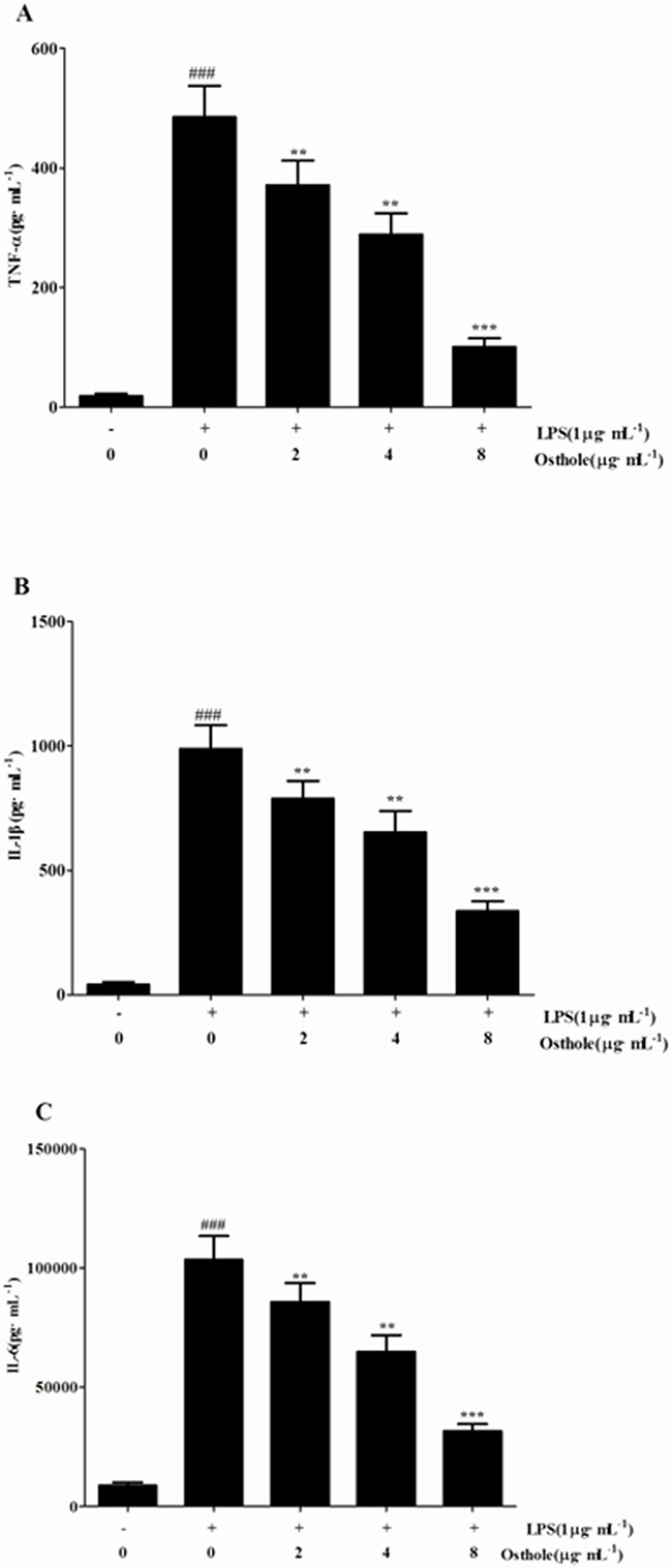
Experimental approach: The protective effect of osthole on mouse macrophage RAW 264.7 and HK-2 cells induced by LPS in vitro and on acute kidney injury model induced by sepsis and established by puncture and cecal ligation (CLP) in vivo were tested.

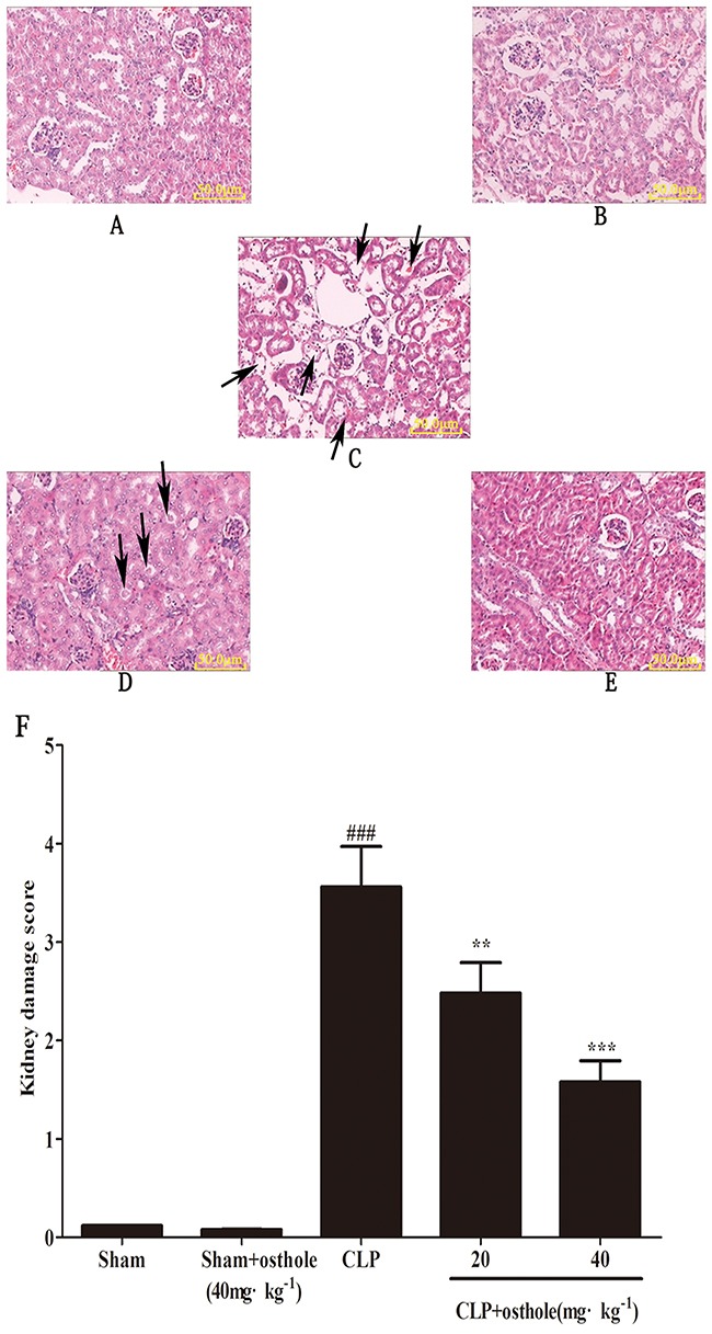
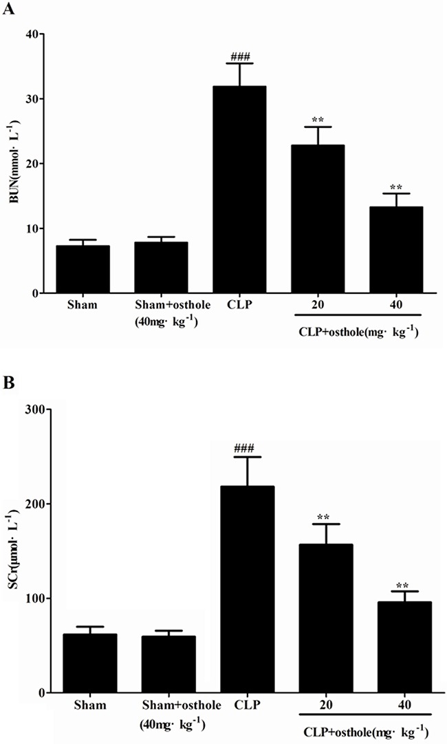
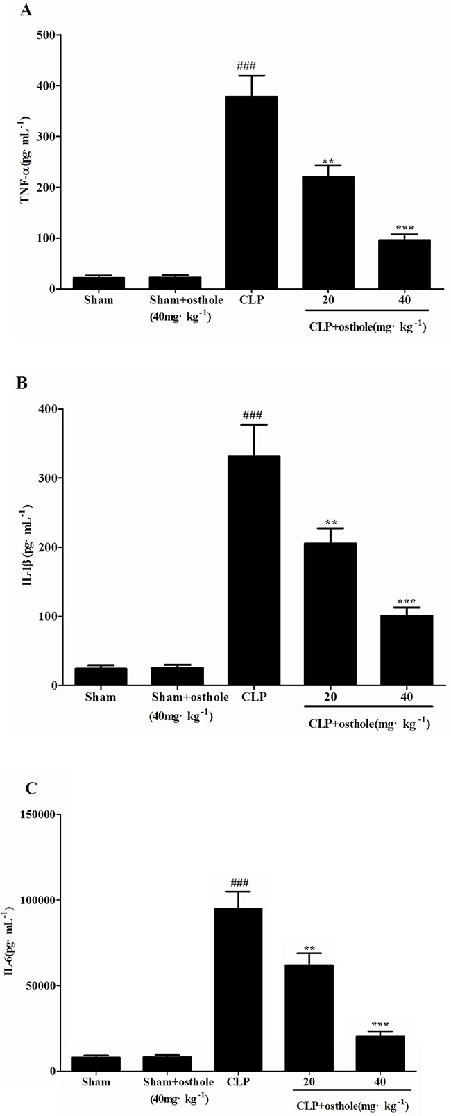
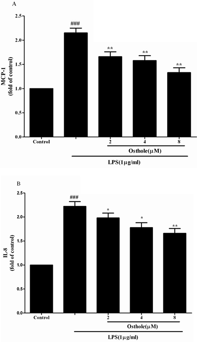
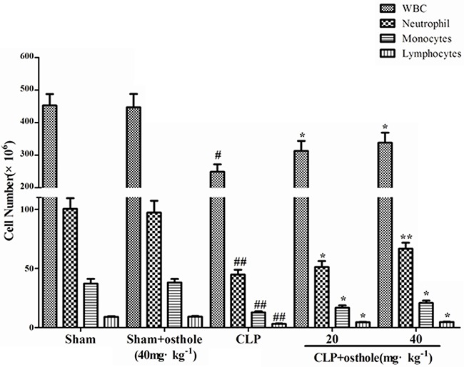
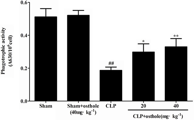
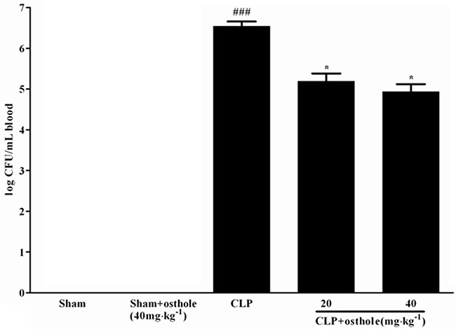
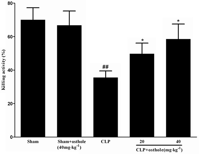
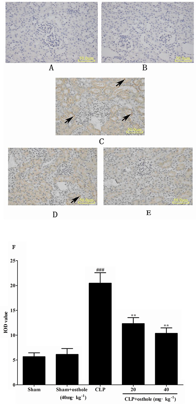
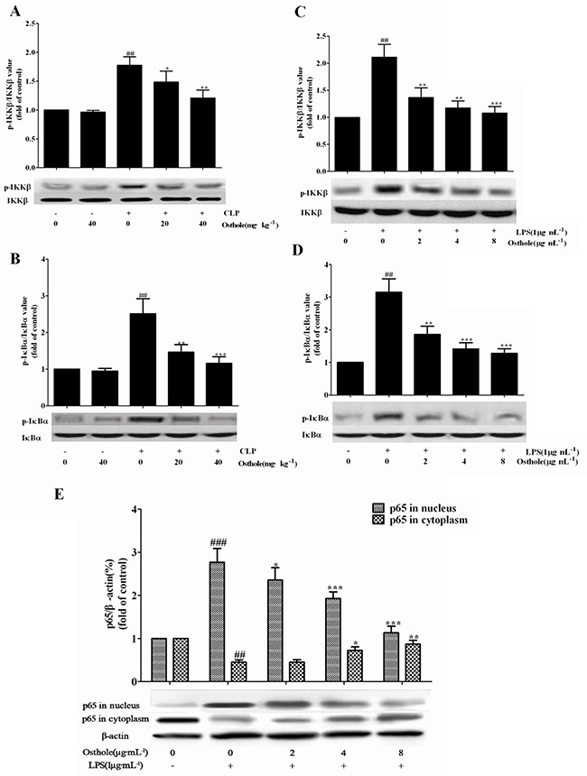
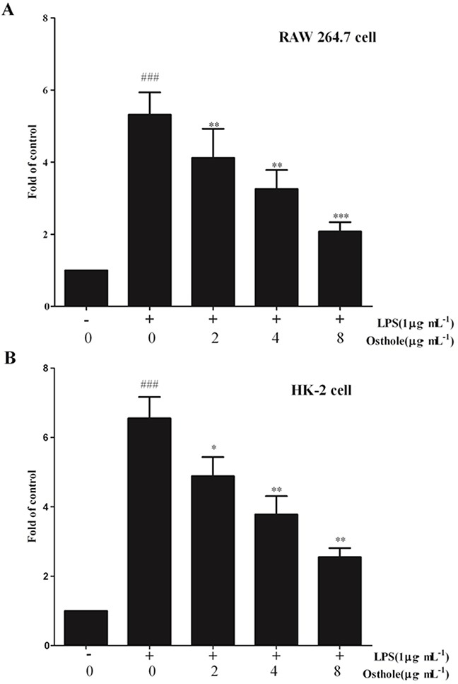
Key results: Osthole (20, 40 mg·kg-1) group can greatly attenuate the changes of the score and kidney histopathology damage and enhance the survival time of septic mice. After the CLP surgery, degrees of SCr and BUN related to kidney injury were upregulated. The concentrations of SCr and BUN can be greatly reduced by treatment with osthole. Furthermore, osthole could increase bacterial killing activity and phagocytic activities of macrophages impaired after CLP partly and attenuate blood bacterial counts and leukocyte infiltration markedly. Furthermore, osthole can suppress NF-κB signal pathway through the inhibition of the nuclear translocation by regulating phosphorylation of IκBα and IKKβ and hinder the production of chemoattractant (MCP-1 and IL-8) and proinflammatory cytokines (TNF-α, IL-1β and IL-6).

Conclusion and implications: Mainly because of its immunomodulatory properties and anti-inflammatory activity, which might be closely associated with suppression of the stimulation of the NF-κB signal pathway, osthole has protective effect on sepsis-induced kidney injury. It can be seen from such evidence that osthole can be potentially applied in the treatment of acute kidney injury.

Keywords: CLP; NF-κB signal pathway; acute kidney injury; osthole; sepsis.


Conflict of interest statement

CONFLICTS OF INTEREST STATEMENT

The authors state that they have no conflict of interest.

Figures

Figure 1
Figure 1. Effect of osthole on survival rate after cecal ligation and puncture (CLP) mice
Mice were challenged by CLP to induce acute kidney injury with or without osthole (20 mg·kg−1 or 40 mg·kg−1) intragastrically administered after operation. Survival was monitored every 6 h during 72 h and the percent survival rate was expressed as Kaplan–Meier survival curves. n=10. ###p< 0.001 compared to Sham group; *p<0.05 and **p<0.01 compared to CLP group.
Figure 2
Figure 2. Effect of osthole on kidney injury after CLP surgery
Representative histological changes in kidneys obtained from mice of different groups A. Sham group; B. Sham+ osthole group; C. CLP group ; D. CLP + osthole (20 mg·kg−1) group; E. CLP + osthole (40 mg·kg−1). The sections shown were harvested 24 h after CLP operation and stained with H&E. Magnification:×100. F. Pathological score of representative kidney samples of each group. The arrowheads indicated the representative morphological changes including tubular degeneration, necrosis and hyperemia. Data are represented as mean ± SD of 10 animals of each group. ###p< 0.001 compared to Sham group, **p<0.01 and ***p<0.001 compared to CLP group.
Figure 3
Figure 3
Effects of osthole on serum BUN A. and SCr B. Data are represented as mean ± SD of 10 animals of each group. ###p< 0.001 compared to Sham group; **p<0.01 compared to CLP group.
Figure 4
Figure 4. Effects of osthole on the production of inflammatory cytokines in serum from mice
Quantitation of TNF-α A. IL-1β B. and IL-6 C. in serum was performed by ELISA. Data are represented as mean ± SD of 10 animals of each group. ###p<0.001 compared to Sham group; **p<0.01 and ***p<0.001 compared to CLP group.
Figure 5
Figure 5. Effects of osthole on the production of inflammatory cytokines by LPS-induced RAW 264
7 cells. Quantitation of TNF-α A. IL-1β B. and IL-6 C. in cultural supernatants was performed by ELISA. Data are represented as mean ± SD of three independent experiments. ###p<0.001 compared to untreated group; **p< 0.01 and ***p<0.001 compared to LPS alone.
Figure 6
Figure 6. Effect of osthole on MCP-1 A. and IL-8 B. release induced by LPS in HK-2 cells
Cells were treated with LPS(1 μg/mL) with or without osthole for 24 h. 100μL of culture medium in each group was taken out to measure the levels of MCP-1 and IL-8 using ELISA kits. Data are representedas mean±SD of three independent experiments. ###p < 0.001 compared to untreated group, *p < 0.05 and **p < 0.01 compared to LPS alone.
Figure 7
Figure 7. Effect of osthole on cells viability tested by MTT assay
A. Effect of osthole on RAW 264.7 cells proliferation in normal condition. B. Effect of osthole on LPS-induced RAW 264.7 cells proliferation. C. Effect of osthole on HK-2 cells proliferation in normal condition. D. Effect of osthole on LPS-induced HK-2 cells proliferation. Results are expressed as percentage of viable cells when compared with control groups. Data are representedas mean±SD of three independent experiments. ##p< 0.01 compared to untreated group; *p< 0.05 and **p< 0.01 compared to LPS alone.
Figure 8
Figure 8. Effect of osthole on peripheral white blood cell counts in CLP-induced sepsis
Blood samples were withdrawn at 24 h after the CLP surgery. Total and differential cell counts were measured. Data are represented as mean ± SD of 10 animals of each group. #p< 0.05 and ##p< 0.01 compared to Sham group; *p< 0.05 and ** p< 0.01 compared to CLP group.
Figure 9
Figure 9. Effect of osthole on peritoneal macrophage phagocytic activity in CLP-induced mice sepsis
Macrophages harvested 24 h after CLP were incubated with zymosan and NBT. Phagocytosis was measured as OD 630 nm. Data are expressed as mean ± SD. (n=10). ##p< 0.01 compared to Sham group; *p< 0.05 and ** p< 0.01 compared to CLP group. NBT: nitroblue tetrazolium.
Figure 10
Figure 10. Effect of osthole on blood bacterial clearance in CLP-induced mice sepsis
CLP animals were orally administrated with osthole at oral dose of 20 and 40 mg·kg−1. Bacteria were counted in blood 24 h after treatment. Colony-forming units (CFU) data are expressed as mean±SD. (n=10). ###p< 0.001 compared to Sham group; * p<0.05 compared to CLP group.
Figure 11
Figure 11. Effect of osthole on bacterial killing activity of macrophages in CLP mice
CLP animals were orally administrated with osthole at oral dose of 20 and 40 mg·kg−1. Peritoneal macrophages were isolated and incubated with E. coli, followed with extensively washing and incubation in fresh medium. Then lysates were got and serially diluted to determine the CFU. Data are expressed as mean±SD. (n=10). ##p<0.01 compared to Sham group; * p<0.05 compared to CLP group. CFU: Colony-forming units.
Figure 12
Figure 12. The effect of osthole on phospho-NF-κB p65 localization and expression in kidney tissue by immunohistochemistry (magnification×100)
A. Sham group; B. Sham+ osthole group; C. CLP group; D. CLP + osthole (20 mg·kg−1) group; E. CLP + osthole (40 mg·kg−1). F. IOD values of phospho-NF-κB p65 staining. Data are represented as mean ± SD of 10 animals of each group, mean IOD values was measured by Image-pro plus 6.0 software. The arrowheads in the stained panels indicate positive staining. ###p< 0.001 compared to Sham group; **p< 0.01 compared to CLP group. IOD: Integral optical density.
Figure 13
Figure 13. Effects of osthole on the activation of the NF-κB signalling pathway
The expression of phospho-IKKβ A. and phospho-IκBα B. in kidneys from CLP-induced AKI mice (n=10), the expression of phospho-IKKβ C. and phospho-IκBα D. and the expression of p65 E. in LPS-induced RAW 264.7 cells were detected by Western blot. Data are represented as mean±SD of three independent experiments. *p< 0.05, ** p< 0.01 and ***p< 0.001 compared to CLP-treated mice or LPS-treated cells ; ## p< 0.01 and ###p< 0.001 compared to sham group mice or untreated cells.
Figure 14
Figure 14. Effect of osthole on the nuclear translocation of NF-κB
A. in RAW 264.7 cell; B. in HK-2 cells. The free NF-κB/p65 in nuclear extracts was assessed using the ELISA-based NF-κB kit 24 h after stimulation. Untreated group is set as 100%. Results are expressed as fold increase over untreated group. Data are represented as mean±SD of three independent experiments. ###p< 0.001 compared to untreated group, *p< 0.05, **p< 0.01 and ***p< 0.001 compared to LPS alone.

References

    1. Lafrance JP, Miller DR. Defining acute kidney injury in database studies: the effects of varying the baseline kidney function assessment period and considering CKD status. Am J Kidney Dis. 2010;56:651–660. - PubMed
    1. Fang Y, Ding X, Zhong Y, Zou J, Teng J, et al. Acute kidney injury in a Chinese hospitalized population. Blood Purif. 2010;30:120–126. - PubMed
    1. Elseviers MM, Lins RL, Van der Niepen P, Hoste E, Malbrain ML, et al. Renal replacement therapy is an independent risk factor for mortality in critically ill patients with acute kidney injury. Crit Care. 2010;14:R221. - PMC - PubMed
    1. Bagshaw SM, Uchino S, Bellomo R, Morimatsu H, Morgera S, et al. Septic acute kidney injury in critically ill patients: clinical characteristics and outcomes. Clin J Am Soc Nephrol. 2007;2:431–439. - PubMed
    1. Murugan R, Karajala-Subramanyam V, Lee M, Yende S, Kong L, et al. Acute kidney injury in non-severe pneumonia is associated with an increased immune response and lower survival. Kidney Int. 2010;77:527–535. - PMC - PubMed

MeSH terms