. 2016 Nov 30;11(11):e0166318.
doi: 10.1371/journal.pone.0166318. eCollection 2016.

Evaluation of the Activity of Lamivudine and Zidovudine against Ebola Virus

Affiliations

Evaluation of the Activity of Lamivudine and Zidovudine against Ebola Virus

Yu Cong et al. PLoS One. .

Abstract

In the fall of 2014, an international news agency reported that patients suffering from Ebola virus disease (EVD) in Liberia were treated successfully with lamivudine, an antiviral drug used to treat human immunodeficiency virus-1 and hepatitis B virus infections. According to the report, 13 out of 15 patients treated with lamivudine survived and were declared free from Ebola virus disease. In this study, the anti-Ebola virus (EBOV) activity of lamivudine and another antiretroviral, zidovudine, were evaluated in a diverse set of cell lines against two variants of wild-type EBOV. Variable assay parameters were assessed to include different multiplicities of infection, lengths of inoculation times, and durations of dosing. At a multiplicity of infection of 1, lamivudine and zidovudine had no effect on EBOV propagation in Vero E6, Hep G2, or HeLa cells, or in primary human monocyte-derived macrophages. At a multiplicity of infection of 0.1, zidovudine demonstrated limited anti-EBOV activity in Huh 7 cells. Under certain conditions, lamivudine had low anti-EBOV activity at the maximum concentration tested (320 μM). However, lamivudine never achieved greater than 30% viral inhibition, and the activity was not consistently reproducible. Combination of lamivudine and zidovudine showed no synergistic antiviral activity. Independently, a set of in vitro experiments testing lamivudine and zidovudine for antiviral activity against an Ebola-enhanced green fluorescent protein reporter virus was performed at the Centers for Disease Control and Prevention. No antiviral activity was observed for either compound. A study evaluating the efficacy of lamivudine in a guinea pig model of EVD found no survival benefit. This lack of benefit was observed despite plasma lamivudine concentrations in guinea pig of about 4 μg/ml obtained in a separately conducted pharmacokinetics study. These studies found no evidence to support the therapeutic use of lamivudine for the treatment of EVD.


Conflict of interest statement

The SRI international affiliation does not alter our adherence to PLOS ONE policies on sharing data and materials.

Figures
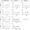
Fig 1
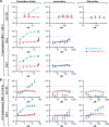
Fig 1. Antiviral activity against Kikwit variant of Ebola virus.
Vero E6, Hep G2, HeLa, or Huh 7 cells were treated 1 h with toremifene citrate (A), lamivudine (B), or zidovudine (C). Two-fold dilutions of the drugs were tested in a 4- to 8-point dose-response curve. Then cells were infected at a multiplicity of infection (MOI) of 1 for 48 h. Toremifene citrate was used as a positive control. Antiviral activity is shown in blue and cytotoxicity is shown in red. The experiment was run on duplicate plates with triplicate wells per dose (mean ± SD; n = 3). Representative graphs from 1 to 4 independent experiments are shown.
Fig 2
Fig 2. Antiviral activity against Makona variant of Ebola virus at an MOI of 1 or 0.1.
(A) Vero E6, HeLa, or Huh 7 cells were treated with toremifene citrate, lamivudine, or zidovudine for 1 h. Then cells were infected with EBOV/Mak at a multiplicity of infection (MOI) of 1 for 48 h. (B) Vero E6 or Huh 7 cells were treated with toremifene citrate, lamivudine, or zidovudine for 24 h. Then cells were infected with EBOV/Mak at a multiplicity of infection (MOI) of 0.1 for 72 h. Toremifene citrate was used as a positive control. Antiviral activity is shown in blue and cytotoxicity is shown in red. The experiment was run on duplicate plates with triplicate wells per dose (mean ± SD; n = 3). Representative graphs from 1 to 6 independent experiments are shown.
Fig 3
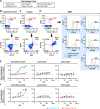
Fig 3. Antiviral activity of lamivudine and zidovudine against EBOV/Mak in MDMs.
(A) MDMs were surface stained with the macrophage panel markers listed. (B) The unstained control sample was stained with LIVE/DEAD Fixable Yellow Dead Cell Stain (Yellow ViD L/D) only. (C, D) The MDM population was measured according to double-positive CD11b and HLA-DR gating. The expression of individual macrophage markers (red line) were gated based on the isotype control (blue line). The percentage of cells positive for each marker is indicated (> 90%). (E, F) MDMs were treated with toremifene citrate, lamivudine, or zidovudine and infected with EBOV/Mak at a multiplicity of infection (MOI) of 0.1. (E) MDMs were pre-treated for 24 h before infection, and the assay endpoint was 48 h post-inoculation. (F) MDMs were pretreated for 1 h before inoculation, and the assay endpoint was 72 h post-inoculation. Toremifene citrate was used as a positive control. Antiviral activity is shown in blue and cytotoxicity is shown in red. The experiment was run on duplicate plates with triplicate wells per dose (mean ± SD; n = 3). Representative graphs from 1 to 4 independent experiments are shown.
Fig 4
Fig 4. Antiviral activity of lamivudine and zidovudine against EBOV-eGFP virus.
Huh 7 cells were pre-treated with imatinib mesylate, lamivudine or zidovudine for 1 h and then inoculated with EBOV-eGFP virus at a multiplicity of infection (MOI) of 0.2. Fluorescence was determined at 48 h post-inoculation. Imatinib mesylate was used as a positive control. Antiviral activity is shown in blue and cytotoxicity is shown in red. The experiment was run with a ten-point, two-fold dilution scheme with quadruplicate wells per dose.
Fig 5
Fig 5. Antiviral activity of drug combinations against Makona variant of Ebola virus.
Dose matrix (6 x 6) evaluation of two drug combinations, lamivudine/toremifene citrate and lamivudine/zidovudine. Huh 7 cells were pre-treated with drug combinations for 1 h and inoculated at a multiplicity of infection (MOI) of 0.1 for 72 h. (A) The heat map of percent response shows the antiviral activity of each combination (100% corresponds to “no activity”). (B) The ΔBliss plot indicates how much a combination effect differs from the additive effect as determined by the Bliss model. The experiment was run once with triplicate wells per dose (mean ± SD; n = 3). ΔBliss = 0 (additive effect); ΔBliss <0 (synergistic effect); ΔBliss >0 (antagonistic effect).
Fig 6
Fig 6. Pharmacokinetic study of lamivudine in guinea pigs.
Plasma concentrations of lamivudine in male and female Hartley guinea pigs after a single oral dose, 60 mg/kg. Each data point represents the mean ± SD of n = 3 guinea pigs except for the 48 h time point in females, n = 2.
Fig 7
Fig 7
In vitro and in vivo evaluation of lamivudine activity against GPA- EBOV (A) Huh 7 cells were pre-treated with toremifene citrate or lamivudine for 1 h. Cells were then inoculated at a multiplicity of infection (MOI) of 0.1 for 48 h. Antiviral activity is shown in blue and cytotoxicity is shown in red. The experiment was run on duplicate plates with triplicate wells per dose (mean ± SD; n = 3). Representative graphs from 2 independent experiments are shown. (B) Percent survival and weight loss of lamivudine-treated guinea pigs infected with guinea pig-adapted-Ebola virus/Mayinga variant (GPA-EBOV/May). Two groups of guinea pigs (n = 6) received oral lamivudine at 20 mg/kg or 60 mg/kg daily from d 3 pre-exposure to d 9 post-exposure. In parallel, the mock group (n = 7) was treated with water. All animals were challenged intraperitoneally (IP) with approximately 1300 pfu of GPA-EBOV/May on d 0.

Similar articles

Cited by

References

    1. Feldmann H, Slenczka W, Klenk HD. Emerging and reemerging of filoviruses. Arch Virol Suppl. 1996;11:77–100. - PubMed
    1. Georges AJ, Baize S, Leroy EM, Georges-Courbot MC. [Ebola virus: what the practitioner needs to know]. Med Trop (Mars). 1998;58(2):177–86. - PubMed
    1. Grard G, Biek R, Tamfum JJ, Fair J, Wolfe N, Formenty P, et al. Emergence of divergent Zaire ebola virus strains in Democratic Republic of the Congo in 2007 and 2008. J Infect Dis. 2011;204 Suppl 3:S776–84. PubMed Central PMCID: PMC3218671. - PMC - PubMed
    1. World Health Organization. Ebola situation report—10 June 2016. Geneva, CH: World Health Organization; 2016. Available: http://apps.who.int/iris/bitstream/10665/208883/1/ebolasitrep_10Jun2016_....
    1. Grygorczuk S, Hermanowska-Szpakowicz T. [Viral hemorrhagic fevers as a biological weapon]. Pol Merkur Lekarski. 2003;14(80):146–9. - PubMed

Publication types