. 2016 Nov 30;11(11):e0167237.
doi: 10.1371/journal.pone.0167237. eCollection 2016.

TLR-2 Recognizes Propionibacterium acnes CAMP Factor 1 from Highly Inflammatory Strains

Affiliations

TLR-2 Recognizes Propionibacterium acnes CAMP Factor 1 from Highly Inflammatory Strains

Coralie Lheure et al. PLoS One. .

Abstract

Background: Propionibacterium acnes (P. acnes) is an anaerobic, Gram-positive bacteria encountered in inflammatory acne lesions, particularly in the pilosebaceous follicle. P. acnes triggers a strong immune response involving keratinocytes, sebocytes and monocytes, the target cells during acne development. Lipoteicoic acid and peptidoglycan induce the inflammatory reaction, but no P. acnes surface protein interacting with Toll-like receptors has been identified. P. acnes surface proteins have been extracted by lithium stripping and shown to induce CXCL8 production by keratinocytes.

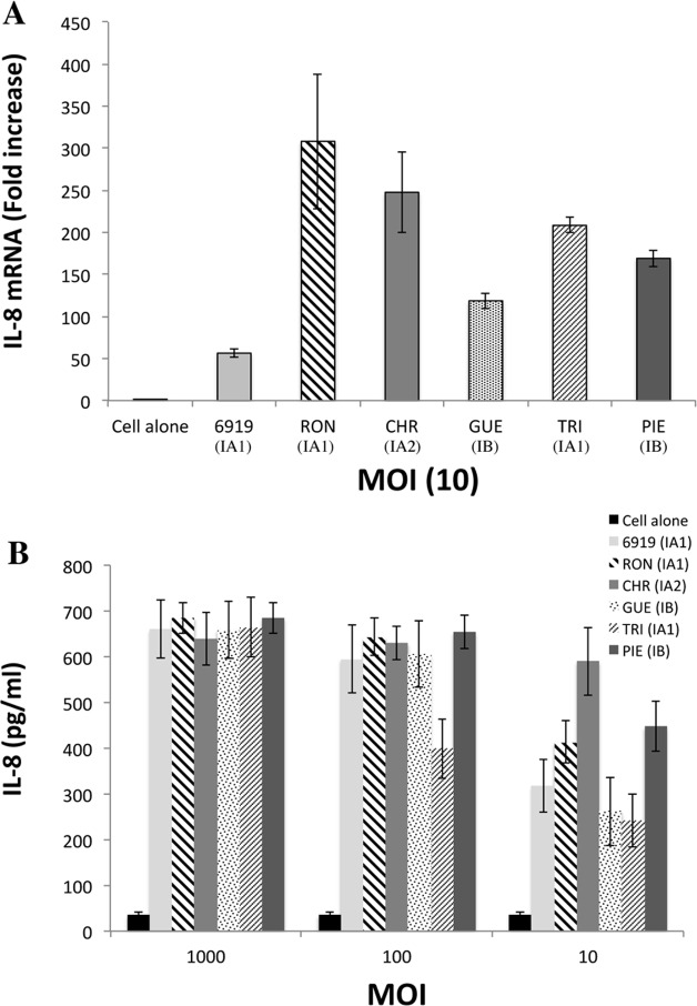
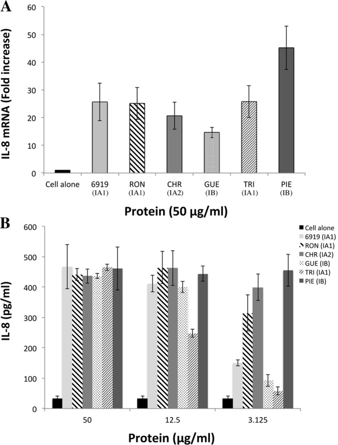
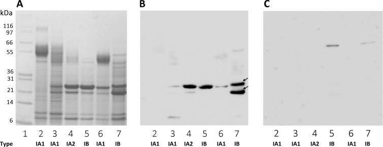
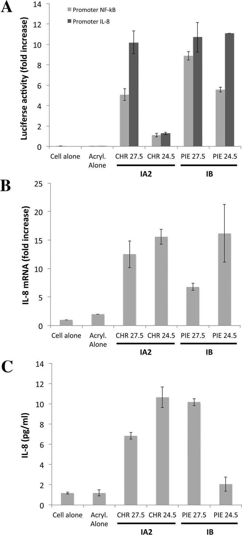
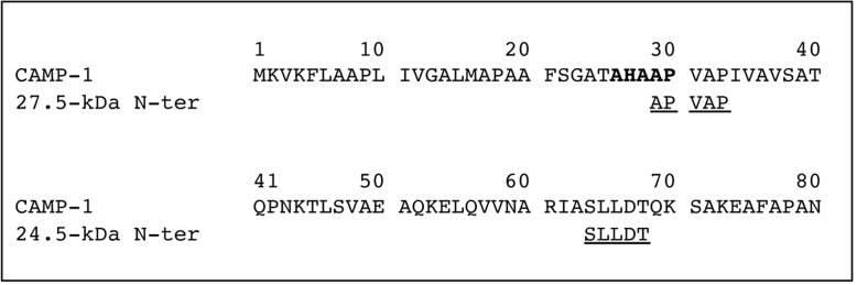
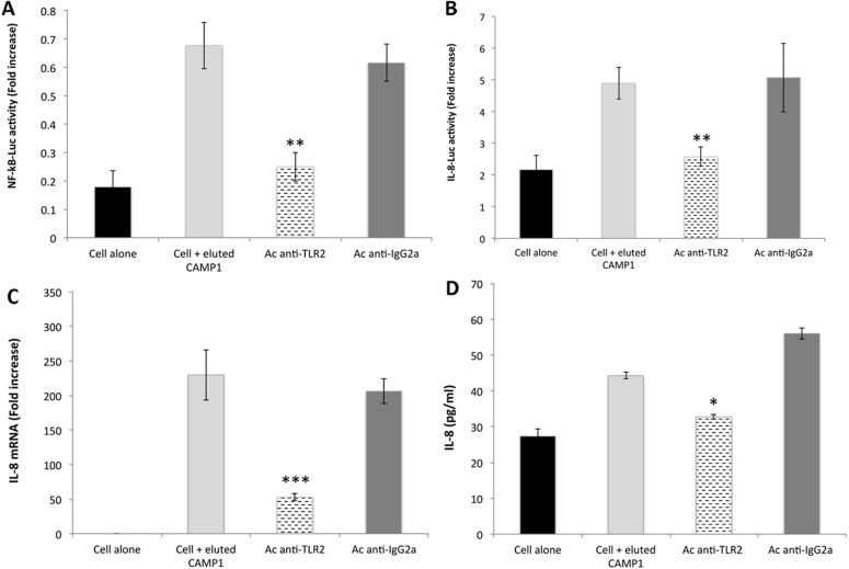
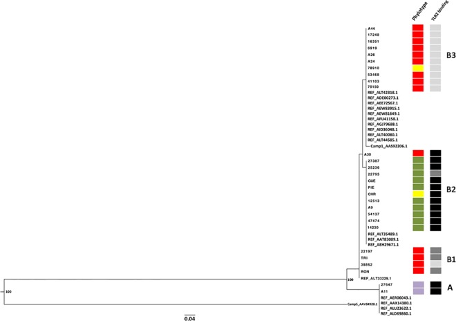
Methodology and principal findings: Far-western blotting identified two surface proteins, of 24.5- and 27.5-kDa in size, specifically recognized by TLR2. These proteins were characterized, by LC-MS/MS, as CAMP factor 1 devoid of its signal peptide sequence, as shown by N-terminal sequencing. Purified CAMP factor 1 induces CXCL8 production by activating the CXCL8 gene promoter, triggering the synthesis of CXCL8 mRNA. Antibodies against TLR2 significantly decreased the CXCL8 response. For the 27 P. acnes strains used in this study, CAMP1-TLR2 binding intensity was modulated and appeared to be strong in type IB and II strains, which produced large amounts of CXCL8, whereas most of the type IA1 and IA2 strains presented little or no CAMP1-TLR2 binding and low levels of CXCL8 production. The nucleotide sequence of CAMP factor displays a major polymorphism, defining two distinct genetic groups corresponding to CAMP factor 1 with 14 amino-acid changes from strains phylotyped II with moderate and high levels of CAMP1-TLR2 binding activity, and CAMP factor 1 containing 0, 1 or 2 amino-acid changes from strains phylotyped IA1, IA2, or IB presenting no, weak or moderate CAMP1-TLR2 binding.

Conclusions: Our findings indicate that CAMP factor 1 may contribute to P. acnes virulence, by amplifying the inflammation reaction through direct interaction with TLR2.


Conflict of interest statement

The authors have declared that no competing interests exist.

Figures

Fig 1
Fig 1. Induction of CXCL8 production in kerationocytes stimulated with whole P. acnes bacteria.
HaCaT cells were incubated for 18 h with whole P. acnes bacteria (6919, RON, CHR, GUE, TRI, PIE, Table 1) at a MOI of 10 (A) and at MOI of 10, 100, 1000 (B). Total RNA was extracted and CXCL8 mRNA levels were determined by real-time RT-PCR and compared those of GAPDH mRNA (used as the control). The difference is expressed as a fold-change. CXCL8 production was analyzed by ELISA on culture supernatants. Control experiments were run with unstimulated cells. The data are presented as the mean ± standard deviation of three independent experiments. Statistical significance is indicated by * P ≤ 0.05, ** P ≤ 0.01, *** P ≤ 0.001, **** P ≤ 0.0001.
Fig 2
Fig 2. Induction of CXCL8 production in keratinocytes by P. acnes surface proteins.
HaCaT cells were incubated for 18 h with lithium extracts of bacterial proteins at concentrations of 50 μg/ml (A) and 3.12, 12.5, and 50 μg/ml (B). Total RNA was extracted and CXCL8 mRNA levels were determined by real-time RT-PCR and compared with GAPDH mRNA levels (used as the control), with expression as a fold-change. CXCL8 production was assessed by ELISA on culture supernatants. Control experiments were performed with unstimulated cells. Data are presented as the mean ± standard deviation of three independent experiments. Statistical significance is indicated by * P ≤ 0.05, ** P ≤ 0.01, *** P ≤ 0.001, **** P ≤ 0.0001.
Fig 3
Fig 3. TLR2 binding to P. acnes surface proteins.
P. acnes surface proteins were extracted from a 5-day culture bacterial pellet and separated by electrophoresis in a 4–12% NuPAGE LDS BisTris gel (50 μg) with detection by Coomassie blue staining (A). Separated proteins were transferred onto nitrocellulose membranes, which were incubated with recombinant TLR2 (B) and TLR4 (C) (0.1 μg/ml). TLR binding activity was detected with specific biotinylated antibodies against TLR2 and TLR4, respectively, as described in the Materials and Methods. Lane 1 contains the molecular mass markers. Lanes 2 to 7 contain proteins from strains 6919, RON, CHR, GUE, TRI, and PIE, respectively. Arrows indicate the positions of the 24.5- and 27.5-kDa bands of interest.
Fig 4
Fig 4. P. acnes surface proteins recognized by TLR2 have proinflammatory activity in vitro.
Surface protein extracts were electrophoretically separated in 10% SDS-PAGE gels (13 x 13 cm), with detection by Coomassie blue staining. Proteins of interest were excised from the gel and eluted as described in the Materials and Methods. (A) HaCaT cells were transfected with CXCL8 (-173 bp) (light gray bar) and NF-κB (dark gray bar) inserted into a construct upstream from the luciferase gene, for 24 h, after which, an internal control (the Renilla luciferase expression plasmid) was added to the transfection mixture. Cells were stimulated by incubation with the eluted proteins (10 μg) for 24 h at 37°C. Relative NF-κB and CXCL8 promoter activities were determined as the ratio of firefly and Renilla luciferase activities. HaCaT cells were stimulated with the eluted proteins (20 μg) for 24 h at 37°C and CXCL8 mRNA levels were evaluated by RT-qPCR (B), and CXCL8 protein levels were assessed by ELISA (C). Data are presented as the mean ± standard deviation of two independent experiments. Statistical significance is indicated by * P ≤ 0.05, ** P ≤ 0.01, *** P ≤ 0.001, **** P ≤ 0.0001.
Fig 5
Fig 5. Comparison of the sequences of the 27.5 kDa P. acnes surface protein and P. acnes CAMP factors.
The peptide sequence of the 27.5 kDa protein was obtained after LC-MS/MS analysis corresponding to 66% coverage (in bold), as described in the Materials and Methods. It was compared with the sequences of P. acnes CAMP factors 1 to 5 (Reference strain NCTC 737, GenBank accession number AY527218.1). Differences between sequences are highlighted in gray.
Fig 6
Fig 6. N-terminal sequencing of the protein of interest.
Surface protein extracts were electrophoretically separated in 10% SDS-PAGE gels (13 x 13 cm), with detection by Coomassie blue staining. Proteins of interest were excised from the gel, eluted and subjected to N-terminal sequencing by Edman degradation to obtain the first five amino-acid residues of each protein, as described in Materials and Methods. N-terminal sequences were compared to the P. acnes CAMP factor 1 sequence (reference stain NCTC 737, GenBank accession number AY527218.1). The peptide signal cleavage site of CAMP factor 1 is shown in bold.
Fig 7
Fig 7. TLR2 blockade inhibits P. acnes CAMP factor 1-induced CXCL8 expression.
Surface protein extracts were electrophoretically separated in 10% SDS-PAGE gels (13 x 13 cm), with detection by Coomassie blue staining. Proteins of interest were excised from the gel, eluted and pooled as described in the Materials and Methods. HaCaT cells were treated for 2 h with human anti-TLR2 (dashed bar) or with goat anti-IgGa antibodies (dark gray bar) and transfected by incubation for 24 h with NF-κB transcription factor (A) and CXCL8 (-173 bp) (B) inserted into a construct upstream from the luciferase gene, after which, an internal control (the Renilla luciferase expression plasmid) was added to the transfection mixture. Cell were stimulated with eluted CAMP factor 1 (50 μg/ml) for 24 h at 37°C. Relative NF-κB and CXCL8 promoter activities were determined as the ratio of firefly and Renilla luciferase activities. Pretreated HaCaT cells were stimulated by incubation with eluted CAMP factor 1 (50 μg/ml) for 24 h at 37°C, CXCL8 mRNA levels were determined by RT-qPCR (C), and CXCL8 production was measured by ELISA (D). Control experiments were performed with cells alone (dark bar) and with stimulated cells not previously treated with CAMP factor 1 (light gray bar). Data are presented as the mean ± standard deviation of two independent experiments. Statistical significance is indicated by * P ≤ 0.05, ** P ≤ 0.01, *** P ≤ 0.001, **** P ≤ 0.0001.
Fig 8
Fig 8. P. acnes CAMP factor 1 polymorphism.
CAMP factor 1 nucleotide sequence analysis was performed on the 27 P. acnes isolates, as described in the Materials and Methods. Nucleotide (dark bold) and peptide (light gray) changes identified in P. acnes isolates relative to the reference sequence of the P. acnes NTCT 737 strain (GenBank accession number AY527218.1) are indicated by vertical bars. (A), (B and C), (D and E), (F) correspond to CAMP1 with no, 1, 2, and several mutations, respectively. See also Table 4.
Fig 9
Fig 9. Phylogenetic analysis of the relationship between P. acnes CAMP factor 1 proteins.
Phylogenetic analysis illustrating the relationship between CAMP factor 1 and 19 protein reference sequences from P. acnes (GenBank accession numbers ALT42318.1, ADE00273.1, AEE72567.1, AEW83915.1, AEW81649.1, AFU41158.1, AGJ79688.1, AID36048.1, ALT40080.1, ALT44585.1, AAS92206.1, ALT35489.1, AAT83098.1, AEH29671.1, ALT33229.1, AER06043.1, AAX14380.1, ALU23622.1, ALD69860.1), CAMP 5 factor Protein (AAV84920.1) and sequences of the 27 P. acnes strains used in this study. Phylogenetic trees were constructed by the maximum likelihood method, using PhyML3.0 [65], and the tree was rooted on the CAMP factor 5 protein sequence. Bootstrapping was applied to the trees (500 datasets) and bootstrap values are shown at nodes. The bar indicates the number of substitutions per site. Sequences were classified into four groups (A, B1 to B3). Squares correspond to phylotype IA1 (red), IA2 (yellow), IB (green), II (purple); and to CAMP1-TLR2 binding intensity with no binding as—(light gray), weak binding as +/- and + (dark gray), strong binding as ++ and +++ (black).

Similar articles

Cited by

References

    1. Grice EA, Segre JA. The skin microbiome. Nat Rev Microbiol. 2011; 9: 244–253. 10.1038/nrmicro2537 - DOI - PMC - PubMed
    1. Aubin GG, Portillo ME, Trampuz A, Corvec S. Propionibacterium acnes, an emerging pathogen: from acne to implant-infections, from phylotype to resistance. Medecine Mal Infect. 2014; 44: 241–250. - PubMed
    1. Horneff JG, Hsu JE, Huffman GR. Propionibacterium acnes infections in shoulder surgery. Orthop Clin North Am. 2014; 45: 515–521. 10.1016/j.ocl.2014.06.004 - DOI - PubMed
    1. Lalani T, Person AK, Hedayati SS, Moore L, Murdoch DR, Hoen B, et al. (2007). Propionibacterium endocarditis: a case series from the International Collaboration on Endocarditis Merged Database and Prospective Cohort Study. Scand J Infect Dis. 2007; 39: 840–848. 10.1080/00365540701367793 - DOI - PubMed
    1. Lomholt HB and Kilian M. Population genetic analysis of Propionibacterium acnes identifies a subpopulation and epidemic clones associated with acne. PLoS ONE. 2010; 5(8): e12277 10.1371/journal.pone.0012277 - DOI - PMC - PubMed

LinkOut - more resources