. 2017 Jan 10;8(2):3072-3103.
doi: 10.18632/oncotarget.13618.

RAC1 GTP-ase signals Wnt-beta-catenin pathway mediated integrin-directed metastasis-associated tumor cell phenotypes in triple negative breast cancers

Affiliations

RAC1 GTP-ase signals Wnt-beta-catenin pathway mediated integrin-directed metastasis-associated tumor cell phenotypes in triple negative breast cancers

Pradip De et al. Oncotarget. .

Abstract

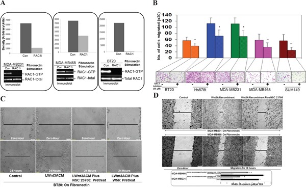
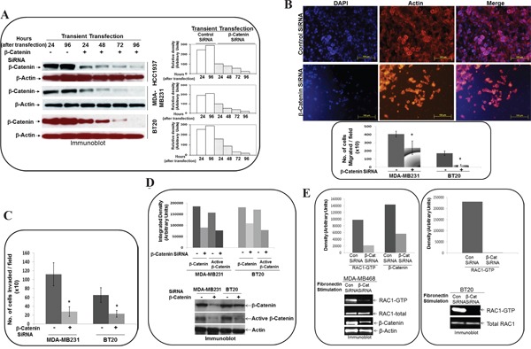
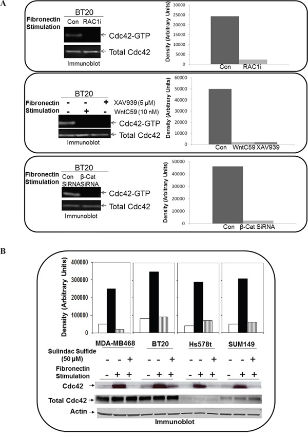
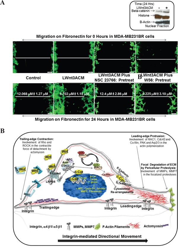
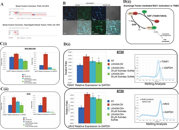
The acquisition of integrin-directed metastasis-associated (ID-MA) phenotypes by Triple-Negative Breast Cancer (TNBC) cells is caused by an upregulation of the Wnt-beta-catenin pathway (WP). We reported that WP is one of the salient genetic features of TNBC. RAC-GTPases, small G-proteins which transduce signals from cell surface proteins including integrins, have been implicated in tumorigenesis and metastasis by their role in essential cellular functions like motility. The collective percentage of alteration(s) in RAC1 in ER+ve BC was lower as compared to ER-ve BC (35% vs 57%) (brca/tcga/pub2015). High expression of RAC1 was associated with poor outcome for RFS with HR=1.48 [CI: 1.15-1.9] p=0.0019 in the Hungarian ER-veBC cohort. Here we examined how WP signals are transduced via RAC1 in the context of ID-MA phenotypes in TNBC. Using pharmacological agents (sulindac sulfide), genetic tools (beta-catenin siRNA), WP modulators (Wnt-C59, XAV939), RAC1 inhibitors (NSC23766, W56) and WP stimulations (LWnt3ACM, Wnt3A recombinant) in a panel of 6-7 TNBC cell lines, we studied fibronectin-directed (1) migration, (2) matrigel invasion, (3) RAC1 and Cdc42 activation, (4) actin dynamics (confocal microscopy) and (5) podia-parameters. An attenuation of WP, which (a) decreased cellular levels of beta-catenin, as well as its nuclear active-form, (b) decreased fibronectin-induced migration, (c) decreased invasion, (d) altered actin dynamics and (e) decreased podia-parameters was successful in blocking fibronectin-mediated RAC1/Cdc42 activity. Both Wnt-antagonists and RAC1 inhibitors blocked fibronectin-induced RAC1 activation and inhibited the fibronectin-induced ID-MA phenotypes following specific WP stimulation by LWnt3ACM as well as Wnt3A recombinant protein. To test a direct involvement of RAC1-activation in WP-mediated ID-MA phenotypes, we stimulated brain-metastasis specific MDA-MB231BR cells with LWnt3ACM. LWnt3ACM-stimulated fibronectin-directed migration was blocked by RAC1 inhibition in MDA-MB231BR cells. In the light of our previous report that WP upregulation causes ID-MA phenotypes in TNBC tumor cells, here we provide the first mechanism based evidence to demonstrate that WP upregulation signals ID-MA tumor cell phenotypes in a RAC1-GTPase dependent manner involving exchange-factors like TIAM1 and VAV2. Our study demonstrates for the first time that beta-catenin-RAC1 cascade signals integrin-directed metastasis-associated tumor cell phenotypes in TNBC.

Keywords: RAC1 GTP-ase; Wnt pathway; actin cytoskeleton; brain-metastasis specific cells; integrin-directed migration.


Conflict of interest statement

CONFLICTS OF INTEREST

The authors declare no conflicts of interest.

Figures

Figure 1
Figure 1. Alterations of RAC1 gene in breast cancers and breast cancer subtypes
A. Oncoprints (cBioPortal) showing alterations in RAC1 gene in Breast Invasive Carcinoma (TCGA Nature 2012) and subtypes including PAM50 Luminal A, PAM50 Luminal B, PAM50 HER2 and PAM50 Basal. The patient selected were, (1) Breast Invasive Carcinoma; (825 patients/825 samples), (2) Breast Invasive Carcinoma, PAM50 Luminal A (235 patients/235 samples), (3) Breast Invasive Carcinoma, PAM50 Luminal B (133 patients/133 samples), (4) Breast Invasive Carcinoma, PAM50 HER2 (58 patients/58 samples) and (5) Breast Invasive Carcinoma, PAM50 Basal (81 patients/81 samples). B. Oncoprints (cBioPortal) showing alterations in RAC1 gene in ER+ve (upper panel) and ER-ve (lower panel) Breast Invasive Carcinoma (TCGA Cell 2015) brca/tcga/pub2015. The oncoprints are generated using 594 patients/594 samples for ER+ve and 174 patients/174 samples for ER-ve breast tumors. C. Oncoprints (cBioPortal) showing alterations in RAC1 gene in Breast Invasive Carcinoma (TCGA Cell 2015) and subtypes including PAM50 Luminal A, PAM50 Luminal B, PAM50 HER2 and PAM50 Basal. The patient selected were, (1) Breast Invasive Carcinoma; (1105 patients/1105 samples), (2) Breast Invasive Carcinoma, PAM50 Luminal A (201 patients/201 samples), (3) Breast Invasive Carcinoma, PAM50 Luminal B (112 patients/112 samples), (4) Breast Invasive Carcinoma, PAM50 HER2 (51 patients/51 samples) and (5) Breast Invasive Carcinoma, PAM50 Basal (107 patients/107 samples). Advanced cancer genomic data visualization is obtained with the help of “The Onco Query Language (OQL)”. Oncoprints (different levels of zoom) have been generated using cBioPortal. Individual genes are represented as rows, and individual cases or patients are represented as columns. Protein level obtained from IHC staining (cBioPortal).
Figure 2
Figure 2. Effect of RAC1 Inhibitors, NSC23766 and W56 on fibronectin-induced RAC1 activation, fibronectin-induced migration, LWnt3ACM stimulated fibronectin-induced migration, and Wnt3A Recombinant stimulated fibronectin-induced migration in TNBC Cells
A. NSC23766 inhibited fibronectin-induced RAC1 activation in MDA-MB231, MDA-MB468, and BT20 cells. TNBC cells were stimulated on fibronectin coated plates. RAC1 pull down was carried out on stimulated cells in the presence and absence of NSC23766. Total RAC1 were used as loading controls. Bar diagram shows changes in the relative density (Arbitrary Units). B. NSC23766 inhibited fibronectin-induced transwell migration in MDA-MB231, MDA-MB468, Hs578t, SUM149 and BT20 cells. Transwell plates were coated with fibronectin and migrated cells were counted following crystal violet staining. Bar diagram (*P< 0.05) shows a number of cells migrated on fibronectin (X20). C. NSC23766 and W56 blocked LWnt3ACM stimulated fibronectin-induced migration in BT20 cells. The scratch assay was performed on fibronectin-coated plates following pretreatment with RAC1 inhibitors. D. NSC23766 blocked Wnt3A Recombinant stimulated fibronectin-induced migration in MDA-MB231 and MDA-MB468 cells. Bar diagram (*P< 0.05) shows changes in the width of scratch (X10).
Figure 3
Figure 3. Effect of WP modulators, WntC59 and XAV939 on fibronectin-induced RAC1 activation and Wnt3A Recombinant stimulated fibronectin-induced migration in TNBC Cells
A. WntC59 and XAV939 blocked fibronectin-induced RAC1 activation in BT20, MDA-MB231 and MDA-MB468 cells. Total RAC1 were used as loading controls. Bar diagram shows changes in the relative density (Arbitrary Units). B. WntC59 and XAV939 blocked Wnt3A Recombinant stimulated fibronectin-induced migration in MDA-MB231 and MDA-MB468 cells. Bar diagram (*P< 0.05) shows changes in the width of scratch (X10).
Figure 4
Figure 4. Effect of WP signaling inhibitor, sulindac sulfide on total beta-catenin and active beta-catenin, fibronectin-induced migration by scratch assay, fibronectin-induced transwell migration, matrigel invasion and Wnt3ARecombinant stimulated fibronectin-induced migration in TNBC cells
A. Sulindac sulfide decreased total beta-catenin as well as transcriptionally active form of beta-catenin in SUM149, MDA-MB231 and BT20 cells. Cells were treated with sulindac sulfide and levels of total and active beta-catenin was determined by Western blots. Actin was used as loading controls. Bar diagram shows changes in the relative density (Arbitrary Units). B. Sulindac sulfide blocked fibronectin-induced migration in SUM149, Hs578t and BT20 cells. Bar diagram (*P< 0.05) shows changes in the width of scratch on fibronectin (X10). C. Sulindac sulfide blocked fibronectin-induced migration in SUM149, MDA-MB231 and BT20 cells. Bar diagram (*P< 0.05) shows a number of cells migrated on fibronectin across the membrane (X20) (upper panel). Migrated cells were stained with phalloidin 555 and counter stained with DAPI. The morphology of the migrated cells was compared with DIC images (lower panel). D. Sulindac sulfide blocked fibronectin-induced matrigel invasion in SUM149 and MDA-MB231 cells. Bar diagram (*P< 0.05) shows a number of cells invaded through matrigel across the membrane (X20). E. Sulindac sulfide blocked fibronectin-induced Wnt3ARecombinant stimulated migration in MDA-MB231 and MDA-MB468 cells. Bar diagram (*P< 0.05) shows changes in the width of scratch as the cell migrate on fibronectin (X10).
Figure 5
Figure 5. Effect of sulindac sulfide on fibronectin-induced cytoskeleton structure of filamentous actin, podia-parameters and activation of RAC1 and Cdc42 in TNBC cells
A-B. Sulindac sulfide altered cytoskeleton structure of filamentous actin in MDA-MB231 and BT20 plated on fibronectin. C-D. Sulindac sulfide blocked lamellipodia while increased filopodia in MDA-MB231 and BT20 plated on fibronectin. Bar diagram (*P< 0.05) shows changes in the numbers of lamellipodia cell migrate on fibronectin. E. Sulindac sulfide abrogated fibronectin induced activation of RAC1 as determined from RAC1-GTP pull-down assay in MDA-MB231, MDA-MB468, BT20, Hs578t and SUM149 cells.
Figure 6
Figure 6. Effect of siRNA-mediated downregulation of beta-catenin on fibronectin-directed migration, matrigel-invasion, transcriptionally active beta-catenin and fibronectin-induced activation of RAC1 in TNBC cells
A. BT20, MDA-MB231 and HCC1937 cells were transiently transfected with beta-catenin siRNA for 96 hours. Total levels of beta-catenin were determined by Western blots and compared between siRNA (24, 48, 72 and 96 hours) and control (24, and 96 hours) cells at similar time points. Actin was used as loading controls. Bar diagram shows changes in the relative density (Arbitrary Units). B. MDA-MB231 and BT20 cells were transiently transfected with beta-catenin siRNA for 24 hours and then allowed to migrate on fibronectin in a transwell assay. Migrated cells were stained with phalloidin 555 and DAPI using the cut-outs of transwell membrane for the picture (upper panel). Bar diagram (*P< 0.05) shows a number of cells migrated through transwell membrane (X10). C. MDA-MB231 and BT20 cells were transiently transfected with beta-catenin siRNA for 24 hours and then allowed to invade across matrigel on fibronectin. Bar diagram (*P< 0.05) shows a number of cells invaded across matrigel (X10). D. Active beta-catenin was determined from lysates of MDA-MB231 and BT20 cells which were transiently transfected with beta-catenin siRNA for 24 hours. Beta-catenin levels were used as the reference for the transient transfection of siRNA. Actin was used as loading control. Bar diagram shows changes in the relative density (Arbitrary Units). E. Active RAC1 (GTP-RAC1) and active beta-catenin were determined from lysates of MDA-MB468, which were transiently transfected with beta-catenin siRNA for 24 hours. Active RAC1 (GTP-RAC1) was determined from lysates of MDA-MB468, which were transiently transfected with beta-catenin siRNA for 24 hours. Actin and total RAC1 were used as loading controls. Bar diagram shows changes in the relative density (Arbitrary Units).
Figure 7
Figure 7. Effect of RAC1 inhibitor, WP modulators, sulindac sulfide and beta-catenin siRNA on fibronectin-mediated activation of Cdc42 in TNBC cells
A. Active Cdc42 was pulled down from BT20 cells treated with NSC 23766 (upper panel), WntC59 and XAV939 (middle panel) and from BT20 cells transiently transfected with beta-catenin siRNA (lower panel). Total Cdc42 were used as loading controls. Bar diagram shows changes in the relative density (Arbitrary Units). B. Active Cdc42 was pulled down from MDA-MB468, BT20, Hs578t and SUM149 cells treated with sulindac sulfide. Actin and total Cdc42 were used as loading controls. Bar diagram shows changes in the relative density (Arbitrary Units).
Figure 8
Figure 8. Effect of RAC1 inhibitors, NSC 23766 and W56 on LWnt3A-stimulated fibronectin-mediated migration of brain metastasis-specific TNBC cells
A. EGFP-tagged brain metastasis-specific TNBC cells, MDA-MB231BR were allowed to migrate on fibronectin for 24 hours under LWnt3A stimulation in the presence or absence of RAC1 inhibitors. Zero hour control for each scratch was presented as the internal control. Activation of the canonical Wnt signaling pathway in MDA-MB231BR cells under LWnt3A stimulation (Inset). Wnt signaling is stimulated in TNBC cells with LWnt3ACM (+) at 24 hours, and is assessed based on the level of beta-catenin accumulation compared to the non-stimulated(-)-condition. Histones were used as a nuclear marker. Beta-actin expression was used as total protein loading control. B. Schematic representation of the mode of involvement of WP via activation of RAC1 in the integrin-mediating directional movement of tumor cells through actin cytoskeletal organization.
Figure 9
Figure 9. Mechanism of RAC1 activation in TNBC following WP stimulation
A. Alterations of TIAM1 and VAV2 genes (exchange factors for RAC1 activation) in breast invasive carcinoma and TNBC subtype:Oncoprints (cBioPortal) showing alterations in TIAM1 and VAV2 genes in Breast Invasive Carcinoma (TCGA, Cell 2015; brca/tcga/pub2015) (upper panel) and TNBC subtype (lower panel). The patient selected were, (1) Breast Invasive Carcinoma; (817 patients/817 samples) and (2) Breast Invasive Carcinoma, TNBC (82 patients/82 samples). B. Activation and nuclear localization of beta-catenin following LWnt3A CM stimulation in MDA-MB231 cells. The White arrow indicates the nuclear localization of beta-catenin (green). DAPI was used for the nuclear staining. The merged picture indicates beta-catenin positive cells. DIC images were captured to test the condition of the cells. C. MDA-MB231BR (i) and BT20 (ii) TNBC cellswere stimulated with LWnt3A CM in the presence and absence of two doses of sulindac sulfide. The mRNA expression of beta-catenin target genes, CCND1 and cMYC were quantified by qRT-PCR. D. Quantification of TIAM1 and VAV2 mRNA by qRT-PCR following sulindac sulfide treatment (25 μM and 50 μM) in BT20 cells under the stimulation of WP by LWnt3A condition media (LWnt3A CM). BT20 (i) TNBC cellswere stimulated with LWnt3A CM in the presence and absence of two doses of sulindac sulfide. The mRNA expression of genes for RAC1 exchange factors, TIAM1 and VAV2 were quantified by qRT-PCR. Melting curves were included in each case. Amplified genomic materials were analyzed for consistency via melting analysis. Each peak represents individual gene products according to base pair size. VAV2 peaks overlap with GAPDH as they are nearly identical in amplicon size. The relative mRNA expression of TIAM1 and VAV2 genes to GAPDH (used as the reference) were determined using LightCycler96 software from Roche. Bars indicate mean ± SE (In triplicates). *P> 0.05. The exchange-factor mediated RAC1 activation in TNBC is schematically represented (ii).
Figure 10
Figure 10. Kaplan-Meier survival for RFS split on the median expression of Affymetrix probe 208640_at RAC1 in IHC determined ER-ve samples in the HAS breast cancer cohort
High expression of RAC1 denotes poor outcome.

References

    1. Entschladen F, TLt Drell, Lang K, Joseph J, Zaenker KS. Tumour-cell migration, invasion, and metastasis: navigation by neurotransmitters. The Lancet Oncology. 2004;5:254–258. - PubMed
    1. Kopfstein L, Christofori G. Metastasis: cell-autonomous mechanisms versus contributions by the tumor microenvironment. Cellular and molecular life sciences. 2006;63:449–468. - PMC - PubMed
    1. Lorusso G, Ruegg C. The tumor microenvironment and its contribution to tumor evolution toward metastasis. Histochemistry and cell biology. 2008;130:1091–1103. - PubMed
    1. Cavallaro U, Christofori G. Cell adhesion in tumor invasion and metastasis: loss of the glue is not enough. Biochimica et biophysica acta. 2001;1552:39–45. - PubMed
    1. Bockhorn M, Jain RK, Munn LL. Active versus passive mechanisms in metastasis: do cancer cells crawl into vessels, or are they pushed? The Lancet Oncology. 2007;8:444–448. - PMC - PubMed

MeSH terms