. 2017 Apr;25(4):533-543.
doi: 10.1016/j.joca.2016.11.008. Epub 2016 Nov 27.

Sex differences in the estrogen-dependent regulation of temporomandibular joint remodeling in altered loading

Affiliations

Sex differences in the estrogen-dependent regulation of temporomandibular joint remodeling in altered loading

J L Robinson et al. Osteoarthritis Cartilage. 2017 Apr.

Abstract

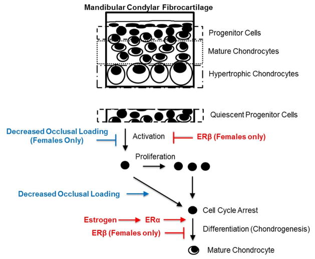
Objective: Temporomandibular joint (TMJ) diseases predominantly afflict women, suggesting a role of estrogen in the disease etiology. Previously, we determined that decreased occlusal loading (DOL) inhibited collagen type II (Col2) expression in the mandibular condylar cartilage (MCC) of female wild-type (WT) mice whereas no change was observed in males. This decrease in chondrogenesis was abolished by estrogen receptor beta (ERβ) deficiency in females. Therefore, the goal of this study was to examine the role of estradiol - ERβ signaling in mediating DOL effects in male mice to further decipher sex differences.

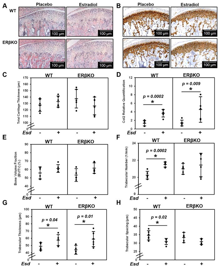
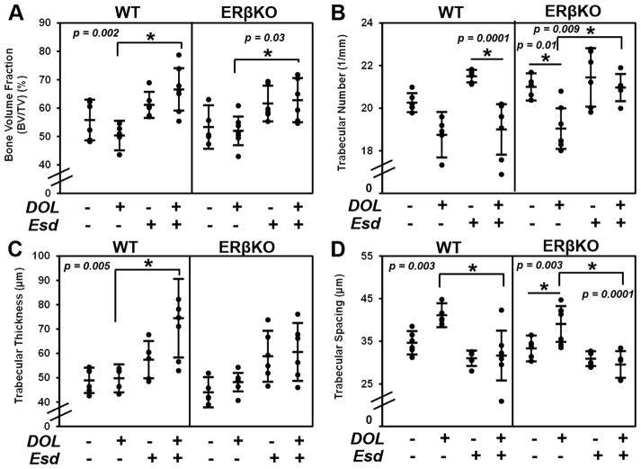
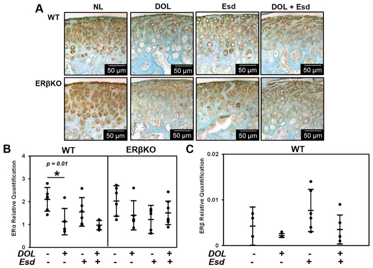
Methods: Male 21 day-old WT and ERβKO male mice were treated with either placebo or estradiol and exposed to normal or DOL for 4 weeks. Cartilage thickness and cell proliferation, gene expression and immunohistochemistry of chondrogenic markers and estrogen receptor alpha (ERα), and analysis of bone histomorphometry via microCT were completed to ascertain the effect of estradiol on DOL effects to the TMJ.

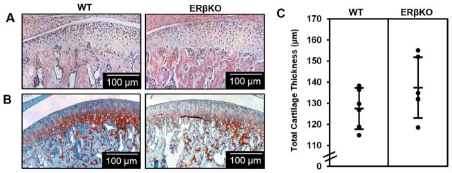
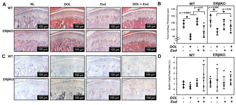
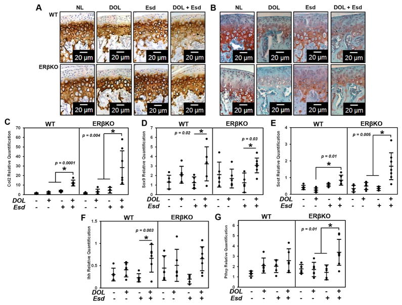
Results: ERβKO male mice lack a MCC phenotype. In both genotypes, estradiol treatment increased Col2 gene expression and trabecular thickness. DOL in combination with estradiol treatment caused a significant increase in Col2 gene expression in both genotypes.

Conclusions: The sex differences in DOL-induced inhibition of Col2 expression do not appear to be mediated by differences in estradiol levels between male and female mice. Greater understanding on the role of estrogen and altered loading are critical in order to decipher the sex dimorphism of TMJ disorders.

Keywords: Decreased occlusal loading; Estradiol; Estrogen receptor β; Mandibular condylar cartilage; Sex differences; Temporomandibular joint.


Conflict of interest statement

9. Conflict of Interest No competing financial interests exist for any of the authors.

Figures

Figure 1
Figure 1. ERβ mandibular condylar cartilage phenotype in male mice
Representative hematoxylin & eosin (H&E) images (A), Safranin-O images (B), and histomorphometric cartilage thicknesses (C) for 49–day WT and ERβKO male mice treated with placebo and subjected to normal load. For histomorphometric analysis, n=6 mice were utilized from each group and the average of 3–6 sections/mouse was analyzed. Statistical significance was determined by a Student’s t-test and p = 0.18.
Figure 2
Figure 2. Effect of estradiol on cartilage thickness, Col2 expression and production, and bone architecture
The data represent WT and ERβKO mice under normal load with either placebo or estradiol treatment. Representative hematoxylin & eosin (H&E) images (A), Col2 immunohistochemical staining (B), cartilage thickness as determined by histomorphometry (C), Col2 gene expression (D), bone volume fraction (E), trabecular number (F), trabecular thickness (G), and trabecular spacing (H) are shown. For histomorphometric analysis, n=6 mice were utilized for all groups and the average of 3–6 sections/mouse was analyzed. For gene expression, n=6 mice were utilized for all groups and mandibular condylar cartilage from left and right were pooled together. Statistical significance was determined by a Student’s t-test and p < 0.05.
Figure 3
Figure 3. Effect of estradiol and decreased occlusal loading on cartilage thickness and cell proliferation
The data represent WT and ERβKO mice under normal load (NL) or decreased occlusal loading (DOL) with either placebo (Plb) or estradiol (Esd) treatment. Specifically, the labels indicate the following: NL = normal load and placebo; DOL = decreased occlusal loading and placebo; Esd = normal load and estradiol; DOL + Esd = decreased occlusal loading and estradiol. Representative hematoxylin & eosin (H&E) images (A), cartilage thickness as determined by histomorphometry (B), BrdU proliferative immunohistochemical staining (C), and quantified percentage of BrdU+ cells (D) are shown. For histomorphometric and BrdU analysis, n=6 mice were utilized for all groups and the average of 3–6 sections/mouse was analyzed. BrdU+ cells were normalized to the total number of cells in the cartilage region as determined from the adjacent H&E section. Statistical significance was determined by a two-way ANOVA followed by posthoc analysis with the Bonferonni method with p < 0.05. Exact p values are listed above the bars that denote significance.
Figure 4
Figure 4. Effect of estradiol and decreased occlusal loading on chondrogenic markers
The data represent WT and ERβKO mice under normal load or decreased occlusal loading with either placebo or estradiol treatment. Specifically, the labels indicate the following: NL = normal load and placebo; DOL = decreased occlusal loading and placebo; Esd = normal load and estradiol; DOL + Esd = decreased occlusal loading and estradiol. Representative Col2 immunohistochemical images (A), representative safranin O images (B) and gene expression of Col2 (C), Sox9 (D), Pthrp (E) and Ihh (F) and Sost (G) are shown. For gene expression, n=6 mice were utilized for all groups and mandibular condylar cartilage from left and right were pooled together. Statistical significance was determined by a two-way ANOVA followed by posthoc analysis with the Bonferonni method with p < 0.05. Exact p values are listed above the bars that denote significance.
Figure 5
Figure 5. Effect of estradiol and decreased occlusal loading on bone architecture
The data represent WT and ERβKO mice under normal load or decreased occlusal loading with either placebo or estradiol treatment. Bone volume fraction (A), trabecular number (B), trabecular thickness (C), and trabecular spacing (D) as determined by microCT are shown. Statistical significance was determined by a two-way ANOVA followed by posthoc analysis with the Bonferonni method with p < 0.05. Exact p values are listed above the bars that denote significance.
Figure 6
Figure 6. Effect of estradiol and decreased occlusal loading on ER- gene expression and immunostaining
The data represent WT and ERβKO mice under normal load or decreased occlusal loading with either placebo or estradiol treatment. Specifically, the labels indicate the following: NL = normal load and placebo; DOL = decreased occlusal loading and placebo; Esd = normal load and estradiol; DOL + Esd = decreased occlusal loading and estradiol. Representative ERα immunohistochemical images (A), and gene expression of ERα (B) and ERβ (C) are shown. For gene expression, n=6 mice were utilized for all groups and mandibular condylar cartilage from left and right were pooled together. Statistical significance was determined by a two-way ANOVA followed by posthoc analysis with the Bonferonni method with p < 0.05. Exact p values are listed above the bars that denote significance.
Figure 7
Figure 7
Working model illustrating the sex-dependent role of estradiol via ERβ and decreased occlusal loading on the mandibular condylar cartilage growth and remodeling.

References

    1. Lipton JA, Ship JA, Larach-Robinson D. Estimated prevalence and distribution of reported orofacial pain in the United States. J Am Dent Assoc. 1993;124:115–121. - PubMed
    1. Manfredini D, Guarda-Nardini L, Winocur E, Piccotti F, Ahlberg J, Lobbezoo F. Research diagnostic criteria for temporomandibular disorders: a systematic review of axis I epidemiologic findings. Oral Surg Oral Med Oral Pathol Oral Radiol Endod. 2011;112:453–462. - PubMed
    1. Tanaka E, Detamore MS, Mercuri LG. Degenerative disorders of the temporomandibular joint: etiology, diagnosis, and treatment. J Dent Res. 2008;87:296–307. - PubMed
    1. Milam SB. Pathogenesis of degenerative temporomandibular joint arthritides. Odontology. 2005;93:7–15. - PubMed
    1. Manfredini D, Arveda N, Guarda-Nardini L, Segù M, Collesano V. Distribution of diagnoses in a population of patients with temporomandibular disorders. Oral Surg Oral Med Oral Pathol Oral Radiol. 2012;114:e35–e41. - PubMed

Publication types

MeSH terms

LinkOut - more resources