. 2016 Dec 19;215(6):875-889.
doi: 10.1083/jcb.201604136. Epub 2016 Nov 30.

NK cells converge lytic granules to promote cytotoxicity and prevent bystander killing

Affiliations

NK cells converge lytic granules to promote cytotoxicity and prevent bystander killing

Hsiang-Ting Hsu et al. J Cell Biol. .

Abstract

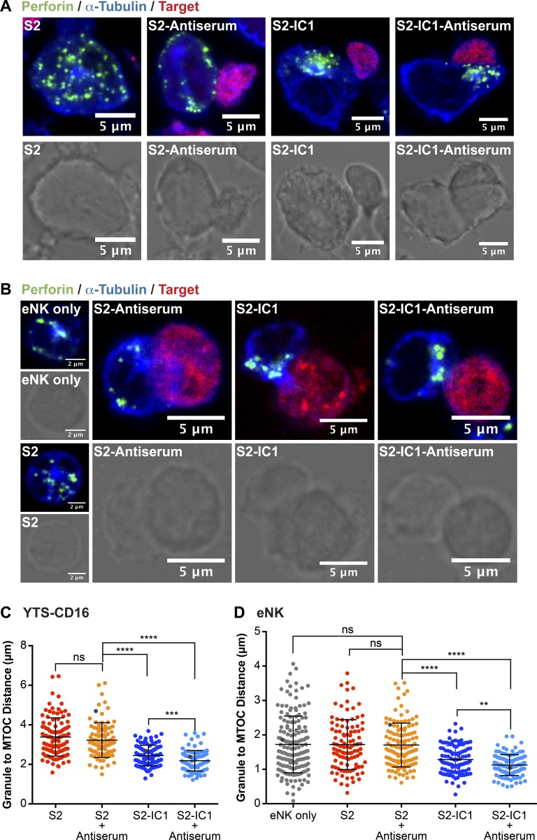
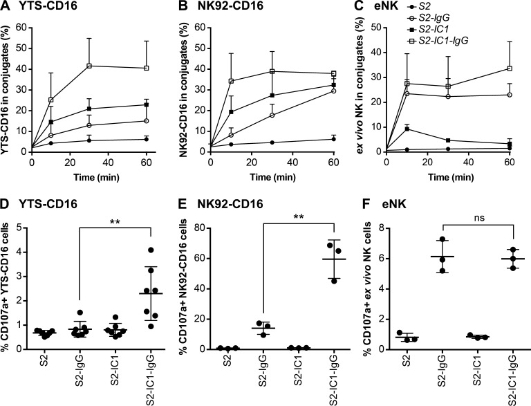
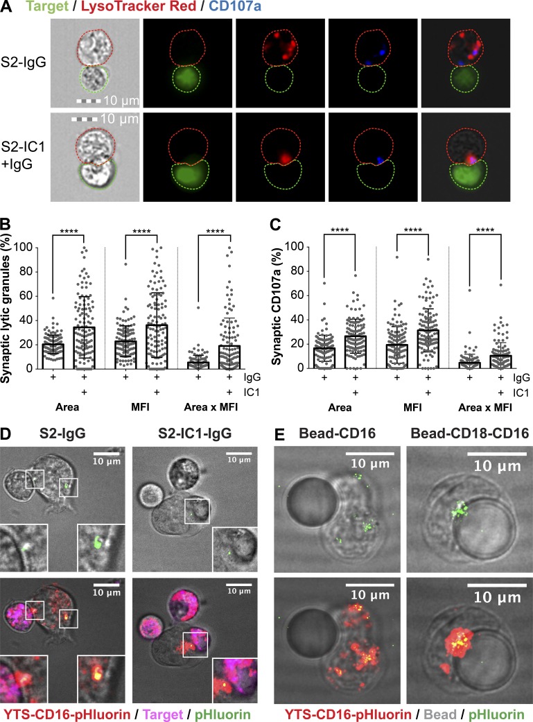
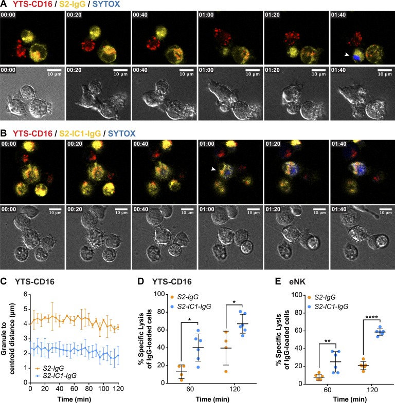
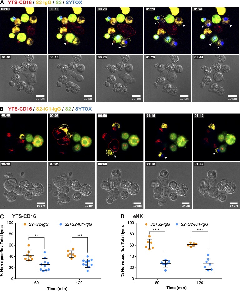
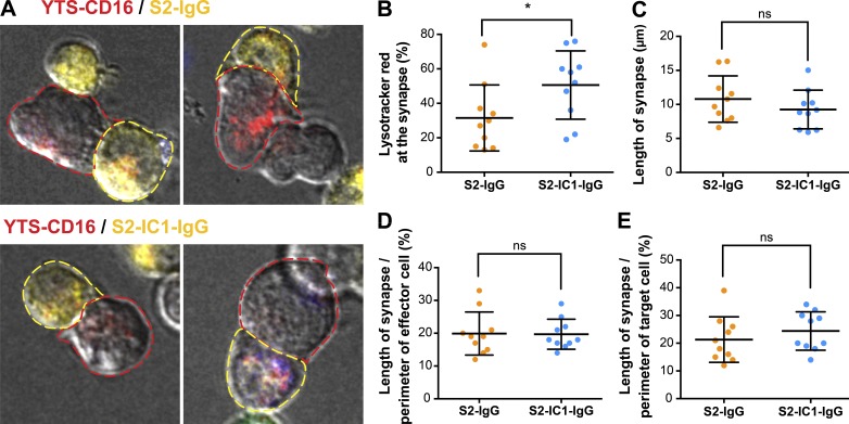
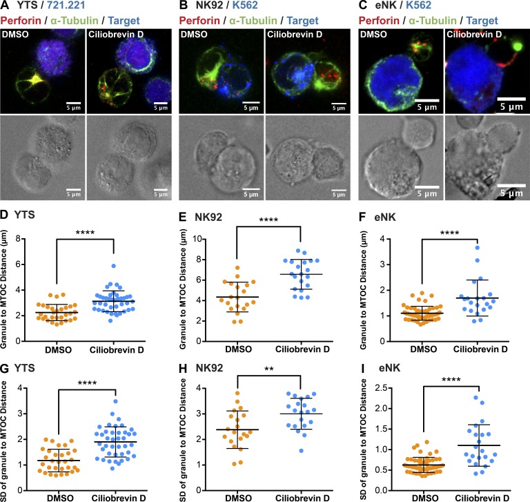
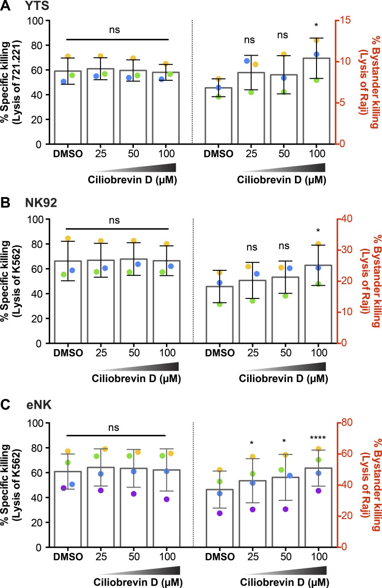
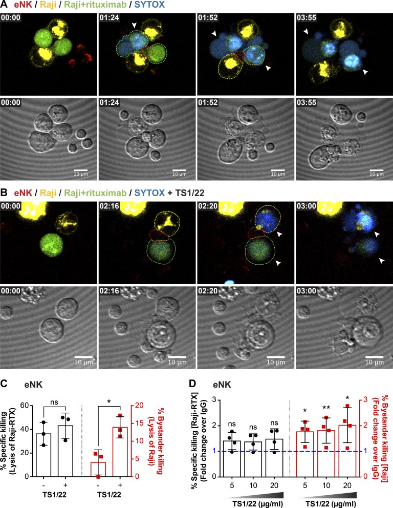
Natural killer (NK) cell activation triggers sequential cellular events leading to destruction of diseased cells. We previously identified lytic granule convergence, a dynein- and integrin signal-dependent movement of lysosome-related organelles to the microtubule-organizing center, as an early step in the cell biological process underlying NK cell cytotoxicity. Why lytic granules converge during NK cell cytotoxicity, however, remains unclear. We experimentally controlled the availability of human ligands to regulate NK cell signaling and promote granule convergence with either directed or nondirected degranulation. By the use of acoustic trap microscopy, we generated specific effector-target cell arrangements to define the impact of the two modes of degranulation. NK cells with converged granules had greater targeted and less nonspecific "bystander" killing. Additionally, NK cells in which dynein was inhibited or integrin blocked under physiological conditions demonstrated increased nondirected degranulation and bystander killing. Thus, NK cells converge lytic granules and thereby improve the efficiency of targeted killing and prevent collateral damage to neighboring healthy cells.


Figures

Figure 1.
Figure 1.
LFA-1 but not CD16 engagement induces lytic granule convergence in NK cells. Fixed-cell confocal microscopy of YTS-CD16 (A) and eNK (B) cells incubated with S2, S2 antiserum, S2-IC1, or S2-IC1 antiserum cells. The NK cells appear by themselves when incubated with uncoated S2, as they did not adhere to the NK cells. Quantitative analyses of lytic granule distance from the MTOC are shown as a feature of the degree of granule convergence in YTS-CD16 (C) and eNK (D) cells. Data represent 30 cells per group, from three independent experiments for YTS-CD16 cells and three healthy donors for eNK cells. Gray points in each condition indicate the representative conjugates shown in A and B. Error bars show ± SD. **, P < 0.01; ***, P < 0.001; ****, P < 0.0001; ns, not significant.
Figure 2.
Figure 2.
CD16 engagement induces conjugate formation and degranulation in human NK cells. For conjugation assay, numbers indicate the percentage of YTS-CD16 cells (A), NK92-CD16 cells (B), and previously cryopreserved eNK cells (C) in conjugates. Data represent results from three independent experiments using NK cell lines or eNK cells from three healthy donors. Error bars indicate + SD (A−C). For degranulation assay, combined results from seven experiments for for YTS-CD16 cells (D) and three experiments for NK92-CD16 cells (E) showed significantly higher degranulation levels of NK cells co-cultured with S2-IC1-IgG cells compared with S2-IgG cells. (F) Data from three healthy donors showed comparable degrees of degranulation by eNK cells coincubated with S2-IgG and S2-IC1-IgG cells. Error bars show ± SD (D−F). **, P < 0.01; ns, not significant.
Figure 3.
Figure 3.
Engagement of LFA-1 and CD16 induces more targeted degranulation at the IS than CD16 alone. (A) Fixed-cell imaging flow cytometry of YTS-CD16 cells conjugated with S2-IgG or S2-IC1-IgG cells. Quantitative analyses of area, mean fluorescence intensity (MFI), and total fluorescence intensity (area × MFI) of LysoTracker red (lytic granules; B) and CD107a (C) staining at the immunological synapse are shown as a feature of directed degranulation of YTS-CD16 cells. Data represent pooled results from three independent experiments; n > 100 cells/group. Error bars show ± SD. Live-cell confocal microscopy of YTS-CD16 cells transduced with a degranulation indicator LAMP1-pHluorin construct conjugated with S2-IgG or S2-IC1-IgG cells (D) or 10 µm polystyrene beads coated with anti-CD16 or anti-CD18 plus anti-CD16 antibody (E). Magenta, target cells; red, LysoTracker red (lytic granules); green, pHluorin (degranulation events). ****, P < 0.0001.
Figure 4.
Figure 4.
Targeted secretion of lytic granules promotes more killing of the target cells. Live-cell confocal microscopy of YTS-CD16 cells conjugated with S2-IgG (A) or S2-IC1-IgG (B) cells. NK cells were mixed with the target cells immediately before the imaging process. Cell mixtures were imaged every 5 min for 2 h. Time zero represents the start of imaging. Yellow, S2-IgG or S2-IC1-IgG cells; red, LysoTracker red (lytic granules); blue, SYTOX blue viability dye. Quantitative analyses of viable cells are shown as a feature of the differential killing efficiency. Arrowheads indicate uptake of SYTOX blue viability dye by the target cells. (C) Live granule tracking in YTS-CD16 cells conjugated with S2-IgG and S2-IC1-IgG cells, respectively. Each point indicates one independent experiment using YTS-CD16 (D) and eNK (E) cells (n > 300 cells/group). Error bars show ± SD. *, P < 0.05; **, P < 0.01; ****, P < 0.0001.
Figure 5.
Figure 5.
Nondirected degranulation outside of the IS increases bystander killing of the neighboring cells. Live-cell confocal microscopy of YTS-CD16 cells incubated with S2 cells as innocent bystanders and S2-IgG (A) or S2-IC1-IgG (B) cells as activating targets. NK cells were mixed with the target cells immediately before the imaging process and imaged every 5 min for 2 h. Yellow, IgG-labeled S2 or S2-IC1 cells; green, bystander S2 cells; red, LysoTracker red (lytic granules); blue, SYTOX blue viability dye. Arrowheads indicate uptake of SYTOX blue viability dye by the target/bystander cells. Quantitative analyses of nonspecific killing of S2 cells over total lysis are shown as a feature of collateral damage to the bystander S2 cells by YTS-CD16 (C) and eNK (D) cells. Each point indicates one independent experiment; n > 400 cells/group. Error bars show ± SD. **, P < 0.01; ***, P < 0.001; ****, P < 0.0001.
Figure 6.
Figure 6.
CD16 ligation alone induces IS geometry similar to that of the IS engaging both LFA-1 and CD16. (A) Example confocal microscopy images of YTS-CD16 cells mixed with differentially labeled S2 cells as performed in Fig. 5. Cell outlines are drawn to indicate the perimeter of the conjugate analyzed (yellow, target; red, effector). Quantitative analyses of the total fluorescence intensity of LysoTracker red at the synapse (B), the length of synapse (C), and the percentage of the perimeter of the cell involved engaged in the synapse from the standpoint of the effector (D) or the target (E). Error bars show ± SD. *, P < 0.05; ns, not significant.
Figure 7.
Figure 7.
Ciliobrevin D inhibits granule convergence in NK cells. Fixed-cell confocal microscopy of YTS (A), NK92 (B), and eNK (C) cells conjugated with their respective target cells after DMSO or ciliobrevin D treatment (100 µM). Red, anti-perforin; blue, 721.221 or K562 cells; green, anti–α-tubulin. Quantitative analyses of the mean lytic granule distance from the MTOC and its SD are shown as a feature of the degree of granule convergence in YTS (D and G), NK92 (E and H), and eNK (F and I) cells. Data represent pooled results from two independent experiments for YTS cells and NK92 cells and two healthy donors for eNK cells (n > 20 cells/group). Error bars show ± SD. **, P < 0.01; ****, P < 0.0001.
Figure 8.
Figure 8.
Ciliobrevin D increases bystander killing of the neighboring cells. Flow cytometry–based cytotoxicity assay of NK cells treated with ciliobrevin D or DMSO control was performed as described in Materials and methods. Raji cells were used as nonsusceptible bystander cells to measure the degree of collateral damage caused by nondirectional degranulation. Specific lysis of the corresponding susceptible targets 721.221 and K562 cells by YTS (A, left) and NK92 (B, left) cells was not affected by ciliobrevin D treatment, whereas the nonspecific lysis of Raji cells by YTS (A, right) and NK92 (B, right) cells increased after ciliobrevin D treatment (100 µM) compared with DMSO control. The cytotoxic function of eNK cells against K562 cells was also not affected (C, left), and the bystander killing of Raji cells was increased (C, right). Data from three independent experiments for YTS and NK92 cells and three healthy donors for eNK cells is shown. Colored dots denote individual experiments or donors. Error bars show ± SD. *, P < 0.05; ****, P < 0.0001; ns, not significant.
Figure 9.
Figure 9.
LFA-1 blockade increases bystander killing. Live-cell confocal microscopy of antibody-dependent cellular cytotoxicity by eNK cells treated with murine IgG1 mAb control (A) or LFA-1 blocking mAb (clone TS1/22; B). Green, RTX-coated Raji cells (NK-inciting targets); yellow, uncoated Raji cells (bystanders); red, LysoTracker red (lytic granules); blue, SYTOX blue viability dye. NK cells were mixed with the target cells immediately before the imaging process and imaged every 4 min for 4 h. Quantitative analyses of viable cells are shown to demonstrate specific (C, left) versus nonspecific (C, right) killing by eNK cells. Data represent combined results from three healthy donors. Standard 4 h 51Cr cytotoxicity assay of NK cells treated with LFA-1–blocking mAb or murine IgG control (D). Each dot represents an individual healthy donor. Error bars show ± SD. *, P < 0.05; **, P < 0.01; ns, not significant.

Similar articles

Cited by

References

    1. Banerjee P.P., Pandey R., Zheng R., Suhoski M.M., Monaco-Shawver L., and Orange J.S.. 2007. Cdc42-interacting protein-4 functionally links actin and microtubule networks at the cytolytic NK cell immunological synapse. J. Exp. Med. 204:2305–2320. 10.1084/jem.20061893 - DOI - PMC - PubMed
    1. Binyamin L., Alpaugh R.K., Hughes T.L., Lutz C.T., Campbell K.S., and Weiner L.M.. 2008. Blocking NK cell inhibitory self-recognition promotes antibody-dependent cellular cytotoxicity in a model of anti-lymphoma therapy. J. Immunol. 180:6392–6401. 10.4049/jimmunol.180.9.6392 - DOI - PMC - PubMed
    1. Blank C., Brown I., Kacha A.K., Markiewicz M.A., and Gajewski T.F.. 2005. ICAM-1 contributes to but is not essential for tumor antigen cross-priming and CD8+ T cell-mediated tumor rejection in vivo. J. Immunol. 174:3416–3420. 10.4049/jimmunol.174.6.3416 - DOI - PubMed
    1. Blott E.J., and Griffiths G.M.. 2002. Secretory lysosomes. Nat. Rev. Mol. Cell Biol. 3:122–131. 10.1038/nrm732 - DOI - PubMed
    1. Bryceson Y.T., March M.E., Barber D.F., Ljunggren H.G., and Long E.O.. 2005. Cytolytic granule polarization and degranulation controlled by different receptors in resting NK cells. J. Exp. Med. 202:1001–1012. 10.1084/jem.20051143 - DOI - PMC - PubMed

MeSH terms